Swell Microscopy
A material found in diapers could change the way large tissue samples are analyzed
What can a highly absorbent polymer found in babys’ diapers do to improve tissue analysis? Researchers from MIT, Cambridge, USA, have used it in their unusual approach to creating high resolution images. “For centuries, a scientist’s ability to look at cells has been constrained by the power of the lenses they used to magnify them. We decided to try something different, and physically magnify the cells themselves,” explains lead author of the associated paper (1), Edward Boyden.
Despite the precision of classical and electron microscopy, Boyden did not find them suitable for the imaging of large, intact 3D tissue samples. “We got frustrated with existing methods of imaging,” he adds “and half-jokingly started talking about just making everything bigger. Then we found papers on swellable polymers that can vastly change their size, and decided to give it a try – the key material in diapers, sodium polyacrylate, is one such polymer, which can absorb a lot of water, swelling enormously in the process.”
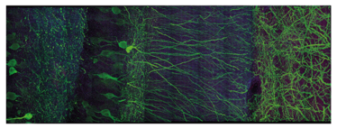
Figure 1. High resolution microscopy of mouse brain tissue following expansion microscopy.
The team were excited to discover that their idea worked in practice. They infused the precursors to this polymer into preserved brain tissue, and then triggered the formation of the polymer chains. The net result: the chains permeate throughout the tissue. “After a few chemical processing steps that make the polymer-embedded tissue very even, we add water, and the polymer chains swell – but because they’re winding their way through the tissue, as they grow, they take the tissue with them, making the tissue itself bigger,” he explains.
Using this technique, which they named expansion microscopy, Boyden and his team were able to expand mouse brain tissue to over four times its original size (Figure 1), without distorting the anatomy, allowing for a more detailed analysis of the morphology. They are now working to increase the expansion factor to allow for even greater magnification. They also hope to adapt other techniques, such as in situ hybridization, in order to identify DNA, RNAs, proteins and biomolecules when analyzing expanded tissues.
- F Chen et al., “Optical imaging. Expansion microscopy”, Science, 347, 543–548 (2015). PMID: 25592419.
I have an extensive academic background in the life sciences, having studied forensic biology and human medical genetics in my time at Strathclyde and Glasgow Universities. My research, data presentation and bioinformatics skills plus my ‘wet lab’ experience have been a superb grounding for my role as an Associate Editor at Texere Publishing. The job allows me to utilize my hard-learned academic skills and experience in my current position within an exciting and contemporary publishing company.


