As a learner and educator in the field of pathology, I have always found comfort in the pinks and blues of pathology images. I remember learning the granulomas of tuberculosis as a medical student by drawing them in different organs – blending histology and pathology without realizing that it was an early, unintentional attempt at integration for better understanding and information recall.
Several years of pathology training and teaching later, on a cruise over the Nile in December 2019, this adventurous educator decided to bring pathology illustrations to life, first on Instagram and expanding soon thereafter to YouTube, LinkedIn and X (formerly Twitter). I found myself opening a door into the microcosm of social media that ties us together in its mysteries and revelations. “Pathodoodles” was born!
The cognitive theory of multimedia learning
Drawing offers many advantages, from comparative histology and summaries to patterns and algorithms. It is not reliant on technology and transcends all generations of learners. Whether using pen and paper or tablets with styluses, drawing unites us through its simplicity of action. No artistic expertise is needed – just a concept in working memory to put down in a drawing. Adding text to the drawing can create concept maps, flow charts, and schematics, which can help summarize a topic and consolidate it concisely. This addresses one of the most critical challenges we face in medical education – cognitive overload.
According to the cognitive theory of multimedia learning, there is a well-understood and proven scientific basis for using illustrations in teaching. When students create illustrations independently during a teaching activity, with the instructor or as a team, they process words and pictures in their working memory, engaging both visual and auditory channels simultaneously. This is far more effective than overloading a single channel, such as the auditory channel, with a passive didactic lecture, or the visual channel with an atlas of images spatially disconnected from the labels or captions.
Once processed, the students integrate the information with their prior knowledge, just like I did – unknowingly – with the granulomas of pathology in year two and the histology of organs from year one. Studies have shown that this learning method significantly enhances students' recall and performance on transfer tests.
Theories aside, learning through illustrations is a delightful and captivating activity for everyone in the classroom. It nurtures a partnership between the instructor and the team, while fostering a sense of personal agency for the student. This approach is a win–win in every aspect of the learning experience and it also caters for every preference, sparking a sense of excitement and curiosity.
The potential for using illustrations in teaching pathology is boundless, as Pathodoodles shows. It is not just about me but about the entire community of pathology enthusiasts. At the time of writing, Pathodoodles has over 250 posts on Instagram with a following of 33.4k people. Pathodoodles also has around 37.6k followers on Facebook and more than 5,600 on X. Many pioneers of pathology education on social media, such as Kamran Mirza and Jerad Garner, have spoken and written extensively about its advantages and potential. #Pathodoodles is just a microscopic – pun intended – part of #PathTwitter and #Studygram. But it is living proof of how we can transcend boundaries using the familiar colors of pathology. And it's all thanks to you, the community.
Pathdoodles’ case studies
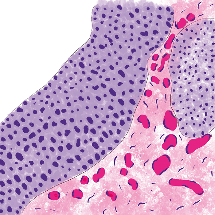
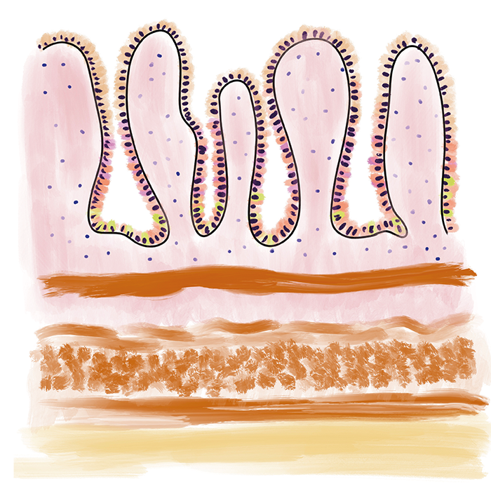
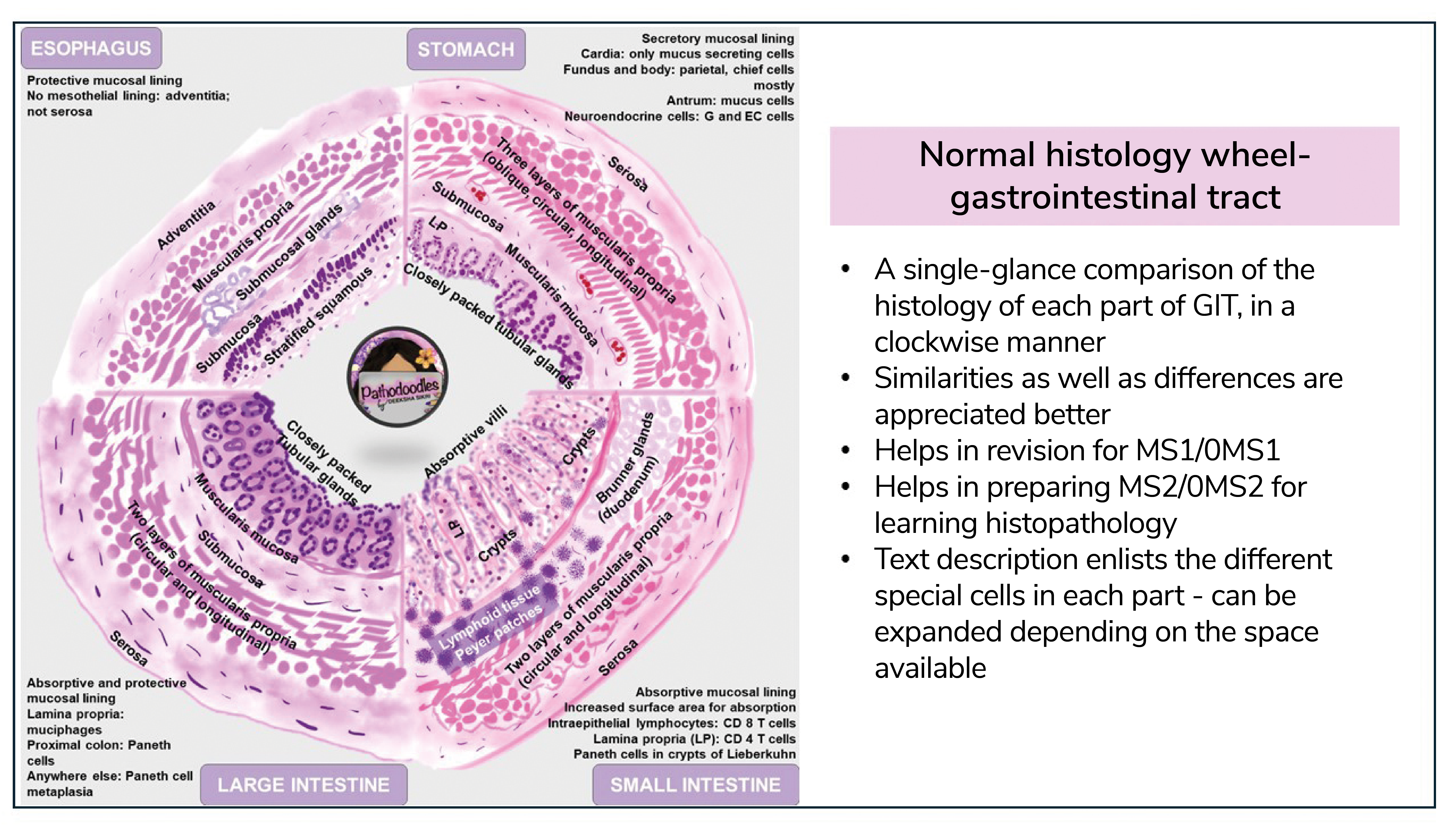
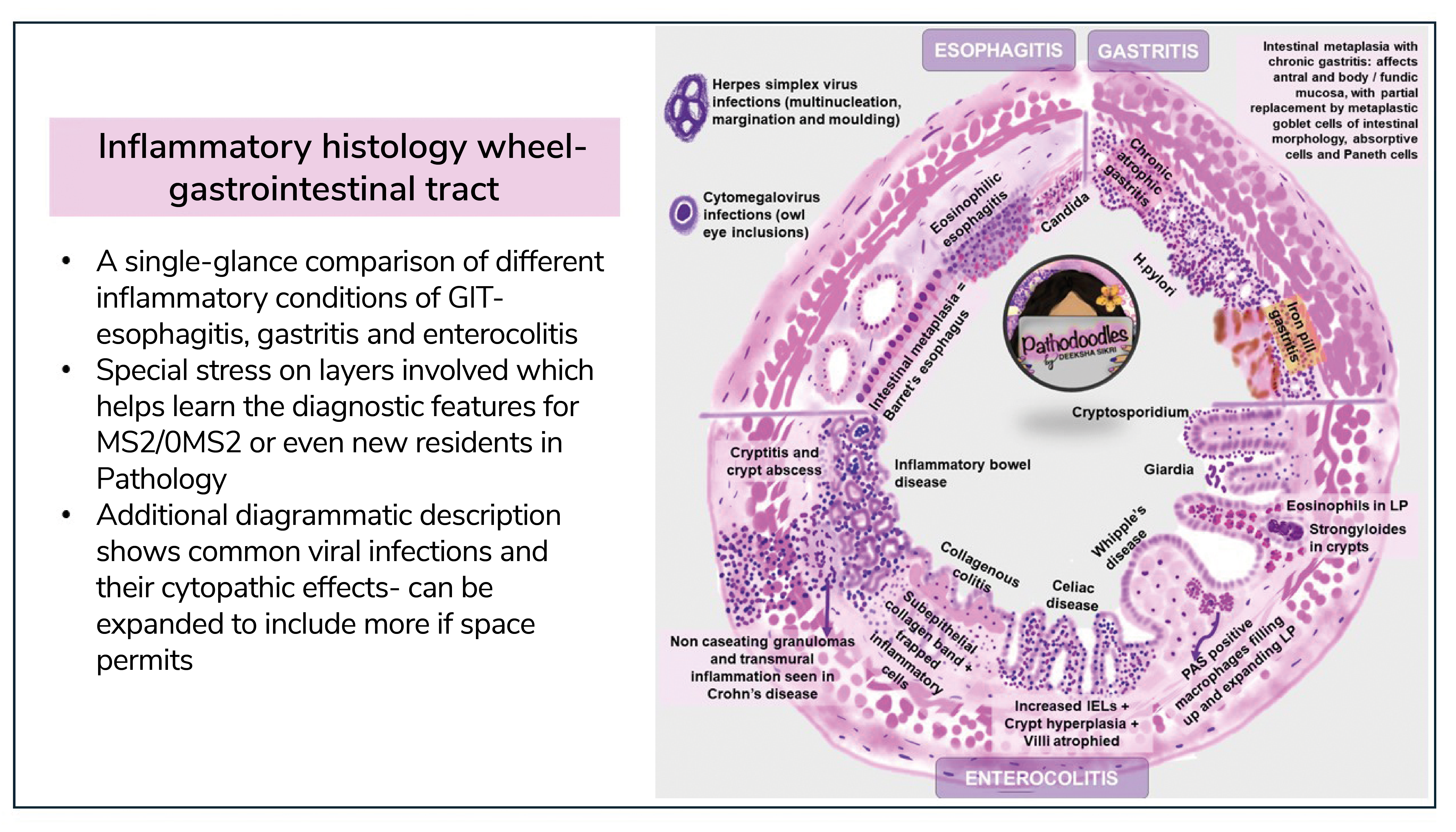
One of the most popular Pathodoodles posts on social media consists of two circular diagrams – one showing normal histology and another depicting inflammatory conditions of the gastrointestinal tract (GIT). This post perfectly illustrates the advantages of using illustrations in pathology education. It supports comparison in two levels: the histology of each part of the GIT compared with each other instantly, and normal histology compared with inflammatory conditions affecting different parts of the GIT. Both wheels are comparisons within themselves and with each other. Normal histology is simplified, and the salient features of each part are highlighted. By presenting these diagrams together the viewer can quickly identify the similarities and differences as you move from the esophagus to the large intestine. The inflammatory pinwheel highlights the layer(s) and cellular infiltrates to help viewers understand and recall the diagnostic features of each inflammatory condition. It also helps to remember the differential diagnosis, as inflammatory conditions have similar clinical pictures.
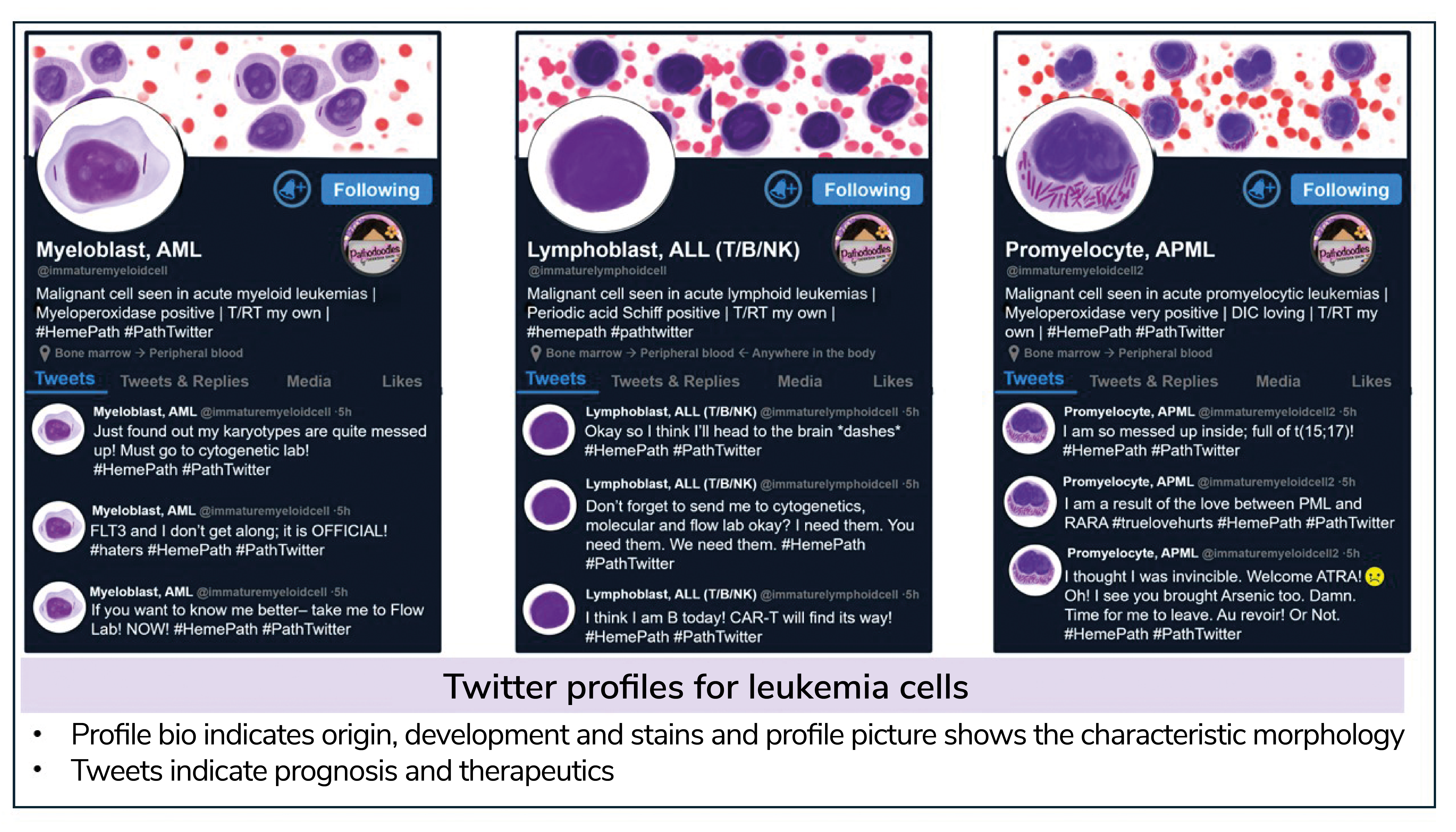
Another popular Pathodoodles approach uses X profiles to explore different leukemia cells. Each profile contains information about the origin of the leukemia cells, the stains they take up, and the genetic changes with prognostic and therapeutic implications. The profile picture shows the morphology of the blast, thus rounding off the salient features of that leukemia. In addition to being an exciting way to learn and recall these leukemias, such illustrations can be used as an assignment or an assessment tool to engage the students more effectively with the learning material.
Pathology’s artistic appeal
Pathology is one of the most artistically appealing branches of medicine. With its many hues and patterns, every section a pathologist views under the microscope every day is a work of art. It is no surprise that the number of a niche kind of pathologist – the “artist” – is rising. The beauty of their art lies not in their accuracy but in their interpretation. With the advent of artificial intelligence, art creation is just a click away. But I believe nothing compares with the mind of an educator and the understanding of what and where their students struggle. Illustrations can bridge gaps in understanding and ease the revision of the vast topics within pathology.
Educational tools used in pathology hold the potential to be used in the field of clinical education as well. For instance, imagine a schematic showing a general pathology concept such as coagulative necrosis – it can easily be extrapolated to cardiovascular pathology showing myocardial infarction, use of cardiac markers in diagnosis, and even treatment options.
Both educators and learners today face challenges of a different kind. The resources available are as abundant as the attention span is short, and this inverse correlation may just get worse as AI use becomes widespread in the field of medical education. Going back to illustrations as a learning and teaching tool is like going back to blackboard teaching – basic, simple, and independent of technology. We only need a pen, paper, and an idea inside the mind of an eager educator and learner.
Image Credit: Pathodoodles
Newsletters
Receive the latest pathologist news, personalities, education, and career development – weekly to your inbox.


