The Multi-omic Cell Atlas
Researchers create a map of human embryonic bone formation
Helen Bristow | | News
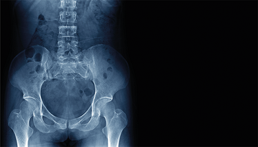
A comprehensive multi-omic study has resulted in a detailed atlas of human embryonic skeletal development, offering insights into the cellular and molecular processes governing bone and joint formation. The research team profiled over 336,000 single nuclei and used spatial transcriptomics to map the development of bones and joints from 5 to 11 post-conception weeks.
By analyzing individual cells, they identified different groups of bone- and cartilage-forming cells and traced two distinct processes: intramembranous ossification, which builds flat bones like the skull, and endochondral ossification, which forms long bones such as those in the limbs. Limb tissues relied on a specific type of cell, called mesenchymal cells, for bone formation, while the skull developed from unique cranial mesenchymal cells.
To better understand the spatial organization of these cells, the team developed a new tool called ISS-Patcher, which maps cell clusters to their precise locations in developing bones and joints. This work revealed key zones within joints and cranial sutures – the areas where bones fuse. In early joints, certain cells favored cartilage formation over bone, guided by the GDF5 gene. In the skull, two important regulators, TWIST1 and RUNX2, were found to control the delicate balance between bone growth and maintaining the flexibility of cranial sutures, shedding light on conditions like craniosynostosis, where these sutures close too early.
The study also explored disease mechanisms using computational tools like SNP2Cell. By linking regulatory networks to genetic variants associated with osteoarthritis and craniosynostosis, researchers predicted cell-specific contributions to these conditions. For example, enhanced RUNX2 activity was linked to hip osteoarthritis, while disruptions to cartilage formation were implicated in knee osteoarthritis.
Corresponding author Sarah Teichmann, said: "Our unique freely available skeletal atlas sheds new light on cartilage, bone, and joint development in the first trimester, detailing the cells and pathways involved together for the first time. This atlas combines cutting-edge spatial technology with genetic analysis and can be used by the research community worldwide.”
Combining my dual backgrounds in science and communications to bring you compelling content in your speciality.


