Finely Tuned Microscopy
Structured illumination microscopy could allow pathologists to “see” glomerular structures, aiding the diagnosis of nephrotic disease
At a Glance
- Structured illumination microscopy (SIM) can help optical techniques overcome their major disadvantage – the diffraction barrier
- Like musicians tune their instruments, microscopists “tune” SIM to obtain clear images of objects much smaller than was previously possible
- SIM has several advantages over the conventional, three-microscope method of diagnosing kidney disease
- Diagnostic technologies in nephropathology – and pathology in general – are always evolving; to stay ahead, pathologists should keep an eye on basic science
Why might a pathologist want to use optical techniques rather than electron microscopy (EM) for diagnosis? Equipment cost, complexity of sample preparation, time investment needed from technicians or pathologists, and even the physical footprint of the device are among the many reasons that exist. Optical imaging can also be highly specific; fluorescent labels can be tagged to specific structures within a specimen, something EM doesn’t make easy. But optical microscopy has one significant failing: resolution. The maximum resolution for an optical microscope, determined by the diffraction barrier, is about 250 nm, whereas an electron microscope can resolve structures a tenth of the size or less – which is why EM has become the technique of choice for diagnosing diseases that require observation of incredibly small features. Nephrotic disease is one such disease family, necessitating the visualization of podocyte foot processes (which are 250–500 nm in size, and can be separated by less than 40 nm) in the glomeruli. But where EM might once have been the only answer, the last 20 years have brought us optical techniques that can overcome the diffraction limit – like structured illumination microscopy (SIM).
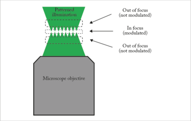
Figure 1. Optical sectioning by SIM.
SIM can beat the diffraction limit by a factor of two, enabling optical resolution down to 100 nm or better. It’s also able to resolve different planes within a sample – “optical sectioning” – letting users visualize a specimen’s three-dimensional structure from a single dataset. Although other new optical techniques can achieve the same or better resolution, SIM has the advantage of being relatively quick and simple. It doesn’t require specialized sample preparation, is compatible with standard fluorescent dyes, and can acquire images through an entire 5 μm thick biopsy section in under five minutes.
How it works
Most microscopy techniques uniformly illuminate the sample, but SIM uses patterned illumination – a periodic array of parallel lines known as a grating. Why? With uniform illumination, light is collected from not just the focal plane, but also other areas, resulting in a blurred background and decreased contrast. With patterned illumination, the grating is formed only in the focal plane of the microscope (see Figure 1); away from the focus, the pattern becomes blurred. That means you can move the grating around within the focal plane to capture different areas, but the background blurring remains constant no matter what. Why is that significant? Because a computer algorithm can play “spot the difference” between several images, eliminating the areas of constant blurring to assemble a sharp image with no background signal.
Another way to look at this process is that the patterned illumination is a form of optical tuning. We “tune” our microscopes using frequency mixing and moiré pattern generation the same way musicians tune their instruments using soundwaves. When tuning the string on a guitar, you chime the tuning fork at the same time as the string and listen to the frequency difference between the two. Then you try to match the string to the tuning fork so that there’s no difference at all.
Just like soundwaves have audible frequencies, we can think of objects and images as containing “spatial frequencies.” Large objects or parts of images correspond to low spatial frequencies, whereas small ones correspond to high spatial frequencies (see Figure 2).
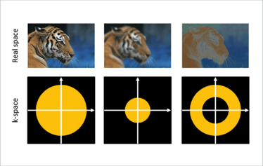
Figure 2. An image (top) contains a range of spatial frequencies, which can be displayed as a k-space map (bottom). Left: full range of spatial frequencies; middle: low frequencies only; right: high frequencies only.
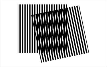
Figure 3. Moiré pattern example. Two high-frequency spatial patterns (vertical lines) intersect to produce a low-frequency pattern (horizontal lines).
One way of expressing the diffraction limit is to say that an optical microscope is only capable of resolving up to a particular spatial frequency. If our grating has a high spatial frequency (close to the diffraction limit), we can use it like an optical tuning fork and probe the spatial frequencies of the sample that were too large for the microscope to observe directly. This produces moiré patterns that the microscope can observe (see Figure 3). If we capture several such images with different grating positions, a computer algorithm can reconstruct an overall image of the sample with higher resolution than a standard microscope can offer.
Renal pathology diagnostics today
At the moment, we diagnose nephrotic disease using a combination of three types of microscopy: EM, immunofluorescence, and conventional light microscopy. This method has its shortcomings, though; EM is time-consuming and requires special tissue preparation, costly equipment, and trained personnel who are becoming increasingly difficult to find. The most significant drawback of all is that the key finding using EM, podocyte foot process effacement, is sometimes difficult to quantitate due to sampling issues – making diagnosis a challenge.
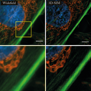
A comparison between standard widefield microscopy (left) and 3D-SIM (right). These bovine pulmonary artery endothelial cells are stained with MitoTracker Red CMXRos (red), Alexa Fluor 488 phalloidin (green), and DAPI (blue). Top: original images; bottom: expanded view of inset.
SIM can help us overcome that obstacle by allowing us to adequately sample and image, over a large area, specimen structures too small to be seen by light microscopy (1). Until recently, that was thought to be impossible, as seeing things below the diffraction limit seemed to violate the laws of physics. In fact, we’re actually cleverly applying those laws to microscopic and mathematical techniques in order to get around the limitations of traditional imaging. SIM requires the use of immunofluorescently labeled proteins, which must be chosen carefully and visualized in darkfield fluorescence mode to generate interpretable images.
How did we come to add this conceptual and technical tour de force to our arsenal of microscopy tools? For that, we owe our thanks to modern laser technologies, high-speed computational techniques, and above all, the intelligence and efforts of the researchers who have worked on these kinds of optical problems for years without recognition. In fact, the 2014 Nobel Prize for Chemistry was awarded for super-resolution microscopy – but unfortunately, SIM pioneer Mats Gustafsson died in 2011 and could not be considered for a share of the prize.
SIM confers several advantages over our current methods:
- It yields high-resolution information that can usually only be obtained by EM.
- It can map specific proteins within tissue at EM-level resolution.
- It can image a much larger volume of tissue at high resolution than is possible with EM.
- Sample preparation is simpler, less expensive, and less time-consuming than it is for EM. Neither preparation nor imaging requires a technician with special training, and the same tissue can be used for both immunofluorescence and SIM.
- The simpler preparation, and the use of light rather than electron beam illumination, appears to create fewer artifacts and less structure distortion than is seen with EM.
- SIM equipment is much smaller and requires less maintenance than an EM setup. High initial equipment cost is an issue at present, but may be addressed with less expensive microscopes in the near future.
SIM in the clinic?
How do we see this all working in practice? We propose that a SIM test for nephrotic disease would be part of the standard kidney biopsy workup in the clinic, done at the same time as conventional immunofluorescence. SIM results would be integrated into the final report, along with immunofluorescence and EM results. Before we can do that, though, we need to devote considerably more time and effort to validating SIM results for clinical use. There are very few published papers and clinical standards have yet to be set up. Would the entrance of SIM into the clinic spell the end of EM’s usefulness? That’s possible in some instances – but SIM technology would need to be much further developed first. Right now, we are at the very dawn of the SIM era.
Ultimately, it’s possible that SIM could be valuable in any area that currently uses diagnostic EM. This includes kidney diseases other than nephrotic syndrome, and might even cover diseases in other organs like the liver, nerves and muscles – although there’s little research using SIM in those areas at the moment. We are primarily working to develop criteria for the SIM-based diagnosis of different types of nephrotic disease. Because SIM presents microscopic structures in a radically different perspective than other types of microscopy, we need to explore further to fully understand what we’re seeing through the microscope. Together, along with our colleagues, we hope to lay down the groundwork for the SIM-based diagnosis of disease.
An eye to basic research
Diagnostic technologies in nephropathology are constantly evolving. Although there are few new ones in the mainstream right now, pathologists and medical laboratory scientists should pay attention to presentations and publications on the applications of basic science to clinical diagnostic problems. Aside from microscopy advances like SIM, there are emerging technologies in molecular genetics and proteomics – for example, mass spectrometry – that are becoming the standard of care in some aspects of renal pathology, like amyloid subtyping. It’s good for clinical professionals to be aware of what’s going on in the world of basic science, because sooner or later, we hope that the best of our new technologies will end up in the clinic, making life easier for the people involved in the diagnosis and monitoring of disease.
Jonathan Nylk is a Research Fellow in the School of Physics and Astronomy at the University of St. Andrew’s, UK.
James Pullman is a Professor of Pathology (Clinical) at Montefiore Medical Center and Albert Einstein College of Medicine, New York, USA.
- JM Pullman et al., “Visualization of podocyte substructure with structured illumination microscopy (SIM): a new approach to nephrotic disease”, Biomed Opt Express, 7, 302–311 (2016). PMID: 26977341.
Jonathan Nylk is a Research Fellow in the School of Physics and Astronomy at the University of St. Andrew’s, UK.
James Pullman is a Professor of Pathology (Clinical) at Montefiore Medical Center and Albert Einstein College of Medicine, New York, USA.


