Exercise Your Diagnostic Acumen
Introducing a collection of slides from the Arkadi M. Rywlin International Pathology Slide Seminar

On behalf of the Arkadi M. Rywlin International Pathology Slide Seminar Organization (IPSSO), I am pleased to make available to the public the proceedings of the 11th Arkadi M. Rywlin International Symposium in Anatomic Pathology, held in Porto, Portugal, May 30–June 1, 2022.
The Arkadi M. Rywlin IPSSO is an international nonprofit organization created in 1990 to foster the exchange of ideas and histopathology slides and to stimulate collegial discussions. The group now comprises more than 50 pathologists from several countries – most of them from Europe and the Americas, but some from farther afield.
The group’s creation was inspired by the legacy of free thinking in pathology fostered by Arkadi M. Rywlin, Professor of Pathology at the University of Miami School of Medicine and Chairman of the Pathology Department at the Mount Sinai Medical Center of Greater Miami. The slide seminar format has allowed the exchange of hundreds of interesting and challenging cases over the years and sparked fascinating discussions, enlightening education and, in many cases, collaborative publications by members of the group.
In 2002, the group decided to expose its activities to a broader audience and held its first live symposium in San Giovanni Rotondo, Italy. Eight years later, Juan Rosai, a founding member of the group, volunteered to scan whole-slide images of every case in the database and make them available to the public through the Juan Rosai Slide Seminar Collection, which he donated to USCAP. Now, thanks to the hard work of many collaborators, we are now able to offer the contents of the last symposium free to the public.
This knowledge-sharing is in memory of a member of our organization – Ondřej Hes. “Ondra” was Professor of Pathology at the Charles University in Plsen, Czech Republic, and an active and enthusiastic member of the Arkadi M. Rywlin IPSSO. Ondra was a preeminent expert in kidney tumors and had likely examined more cases of renal cell carcinoma than anyone else in the world. His expertise was unparalleled and his many contributions to the literature helped shape the landscape of renal tumoral pathology as we know it today. His kindness and generosity always set him apart as a person, and his departure has sent shock waves throughout the system. We mourn his departure as the greatest of losses; our specialty and our lives will be poorer without him. Many great pathologists are remembered for their contributions, but Ondra was truly beloved by everyone he knew. He will always be in our hearts.
I would also like to make public my gratitude to Michael Schubert of The Pathologist for generously agreeing to post the cases of this symposium, to Pathcore for providing a platform to accomplish this, and to Ivan Damjanov, one of our club members (and the person who initially proposed this idea and pursued it to completion). I would also like to thank Manuel Sobrinho-Simões and Fátima Carneiro, from IPATIMUP, University of Porto, for organizing and hosting this event – and all the members of their team, in particular Elisabete Rios, without whose enthusiastic assistance none of this would have been possible. Finally, I would like to thank all the members of the Arkadi M. Rywlin IPSSO for their contributions to this effort. It has only been through their active, selfless participation and support that this organization has flourished over the years.
Saul Suster
Montclair, New Jersey, USA
Click on the thumbnail images below to access the interactive whole-slide images for each case. For the detailed presentation, analysis, and references for each case, please download the case handout.
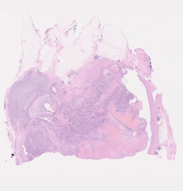
1. A 68-year-old retired nurse presented with a large palpable left breast mass that measured 5 cm by ultrasound. It was biopsied and excised. The slides are from the excision.
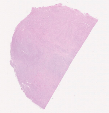
2. Woman, 68 years old. No previous oncological history.
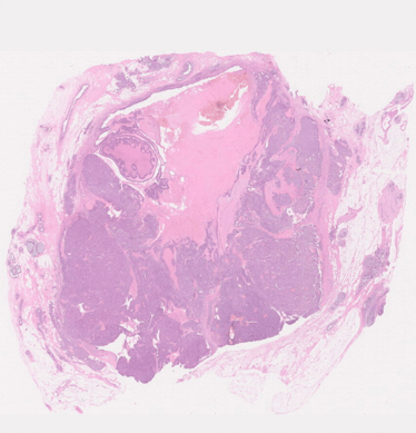
3. A 68-year-old female palpated a right breast mass that was biopsied and excised by mastectomy. The slides are from the mastectomy specimen.
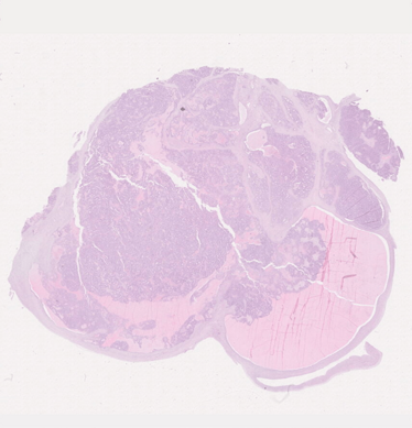
4. A 56 year-old female, status post-left salpingo-oophorectomy, presented to the emergency room with acute urinary retention. CT scan showed a 12 cm right pelvic mass. Her serum CA125 was 1,104 U/mL. She underwent a TAHRS and staging.
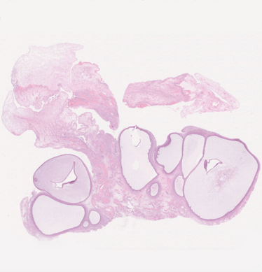
5. A 21-year-old female with abdominal pain and distension of two months. Abdominal CT with ovarian mass. CA125: 244. Ascites and pleural effusion. Resection of abdominal mass and frozen section was performed. Rest of pelvic cavity contained adhesions.
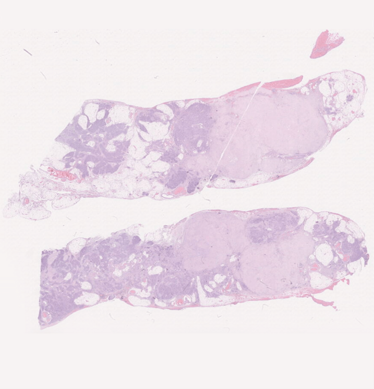
6. An 80-year-old woman with peritoneal tumor deposits and a 10.5 cm partly cystic partly solid right ovarian mass. Submitted slides for the symposium were cut from the metastatic tumor deposits on the peritoneum. A previous peritoneal core needle biopsy showed metastatic adenocarcinoma with focal adenoid-cystic-like features. Hysterectomy, bilateral salpingo-oophorectomy, and omentectomy were performed.
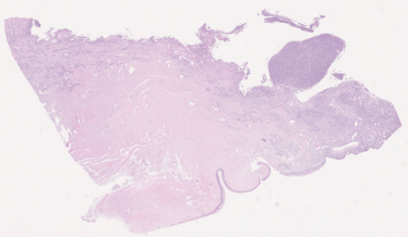
7. A 46-year-old 1G1P female presented with a month history of abnormal vaginal bleeding. Physical examination revealed a 4 cm exophytic friable mass in the uterine cervix. A cervical biopsy was performed and the initial pathologic diagnosis was endometrioid adenocarcinoma. She underwent a total abdominal hysterectomy, bilateral salpingo-oophorectomy, pelvic lymphadenectomy, and omentectomy. The patient is alive with no evidence of disease at 10 months after the surgery.
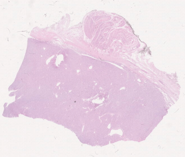
8. A 75 year old female with history of non-Hodgkin lymphoma since 1994 in remission. During follow-up, 24 years after initial diagnosis of lymphoma, she presented with abdominal discomfort and a thoraco-abdominal CT scan showed a pelvic mass, apparently in uterine ligament. Tumor markers were negative. Resection of abdominal mass and frozen section was performed. During surgery, abdominal cavity was clear of disease.
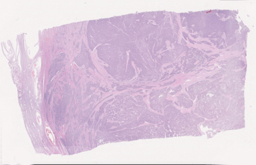
9. A 40-year-old woman without significant clinical history presented with abnormal uterine bleeding. Curetting was performed, followed by total abdominal hysterectomy. A 4.9 cm well-circumscribed but not encapsulated nodule was seen within the myometrium. The cut surface was homogeneous whitish to tan. No additional nodules were noted. At the time of surgery, no metastases were detected. The patient was lost to follow-up.
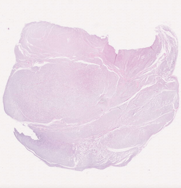
10. A 42-year-old female patient presented with a well-circumscribed soft lesion at the vulva that was completely excised.
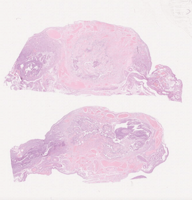
11. A 38-year old female presented with lower abdominal discomfort. Imaging analyses indicated multiple uterine myomas. A bilateral salpingectomy and myomectomy were laparoscopically performed. The patient is fine and free from disease 36 months after surgery.

12. A 46 year-old woman presented with a five-month history of abdominal pain. CT scan of abdomen and pelvis showed a 20 cm multicystic right pelvic mass and mild to moderate ascites. Her serum CA125 was 386 U/mL. Her personal history was unremarkable; family history showed that her father had skin and prostate cancer in his late 60s. The patient underwent TAHBSO, omentectomy, pelvic and periaortic lymph node sampling, and pelvic washings. She also received six cycles of adjuvant chemotherapy with carboplatin and paclitaxel. A blood sample was submitted for hereditary ovarian cancer panel germline testing that included BRCA1, BRCA2, BRIP1, EPCAM, MLH1, MSH2, MSH6, PALB2, PMS2, RAD51C, and RAD51D. No pathogenic mutations were identified.
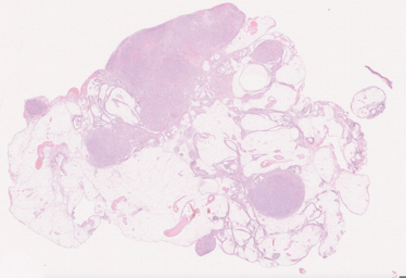
13. A 69 year-old, 4G3P female was admitted because of ascites and an enlarged right ovary. Clinical and image studies indicated a right ovarian cancer, stage IV (lung and peritoneal wall metastases). The patient was treated with simple abdominal hysterectomy, bilateral salpingo-oophorectomy, and omentectomy with preoperative chemotherapy. Intraoperative cytologic and histological consultation was performed. Serum alpha-feto protein (AFP) levels were elevated (up to 269 ng/mL) after surgery. The patient died of disease eight months after surgery.
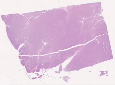
14. An eight-year-old girl presented with a 14 cm retroperitoneal mass. Resection was performed. The slides are from this resection specimen.
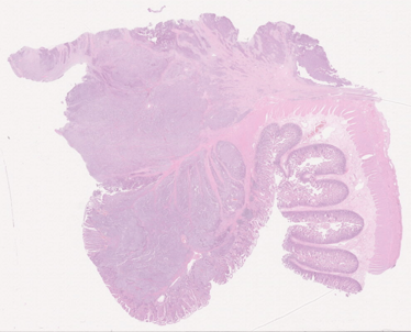
15. A 17-year-old male with a 6.6 cm mass of proximal ileum resected.
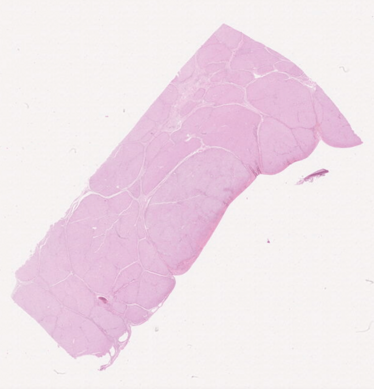
16. This nine-year-old boy presented with abdominal discomfort and swelling. His abdominal symptoms and hospitalizations dated back to when he was seven months old; in the workup at that time, he was diagnosed with tyrosinemia. His parents are second-degree relatives. Radiologic examination revealed liver nodularity, some with characteristics of hepatocellular carcinoma. AFP was 125,000. Albumin was 3.5 g/dL, ALP 99 (low), bilirubin 0.2 (low), INR 1.03 (high). After detailed workup, the patient underwent liver transplant. The specimen you see is from the explant.
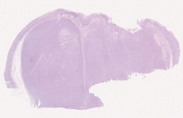
17. An 18-year-old female with two gastric masses, 10 and 3 cm, and extensive serosal/peritoneal nodules. Lymph nodes were also involved.
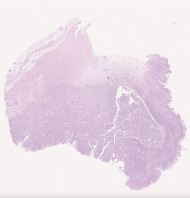
18. Male, 86 years old, total gastrectomy with a polypoid tumor (4.6cm in largest dimension) localized to the esophago-gastric region.
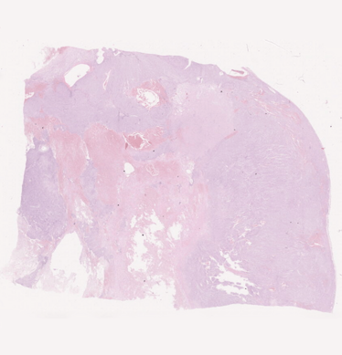
19. A 50-year-old male with an abdominal pelvic mass excised, about 8 cm in greatest diameter. Tumor was whitish, rubbery, solid, and homogeneous.
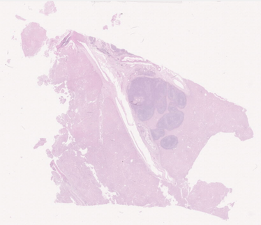
20. Man, 71 years old. No previous oncological history. US performed during a checkup for back pain revealed a 2.2 cm lesion in the liver. Imaging (PET; CT; MNR) negative for other lesions. No biopsy performed. Preoperative diagnosis: cholangiocarcinoma.
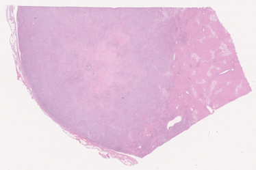
21. This 61-year-old male patient presented with abdominal pain and was discovered to have multiple tumors in the liver, the largest measuring 8 cm. After a biopsy, the patient underwent liver transplantation. The specimens you see are from the explant.
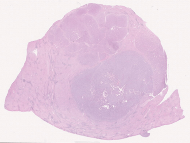
22. A 67-year-old man with a clinical history of T cell acute lymphoblastic lymphoma, and hepatitis C was found to have a 2 cm mass in the left lobe of the liver on routine surveillance imaging.
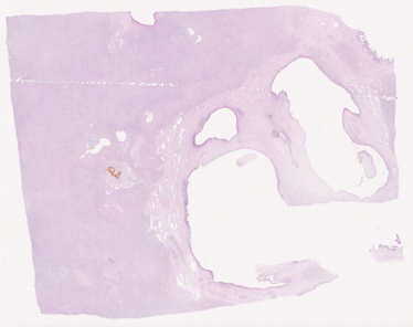
23. Female, 24 years old. Liver specimen (partial hepatectomy) with a large tumor (13.5 cm in largest dimension) with necrotic areas.
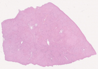
24. A 38-year-old man with a history of interstitial lung disease and combined variable immunodeficiency (CVID) presented with severe sequelae of portal hypertension, including massive splenomegaly (28.4 cm spleen on imaging), and splenorenal, retroperitoneal, and gastroesophageal varices. Liver function tests were mildly but chronically elevated, previously thought to be due to non-alcoholic fatty liver disease because the patient was obese.
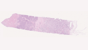
25. Man, 74 years old. History of benign prostatic hyperplasia (TURP performed elsewhere). US performed during a checkup for acute urine retention revealed three lesions in the liver. Imaging (PET; CT; MNR) negative for other lesions. No biopsy performed. Preoperative diagnosis: cholangiocarcinoma.
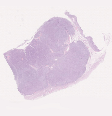
26. Male, 77 years old, total gastrectomy with an ulcerated tumor (4.5cm in largest dimension) localized in the gastric body.
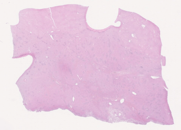
27. A 41-year-old woman with multiple large liver masses that grew steadily over a two-year period. Despite conservative management and discontinuation of oral contraceptives for two years, transplantation was required due to disease burden. The patient was found to have a mutation in PALB2 (a BRCA2 partner gene).
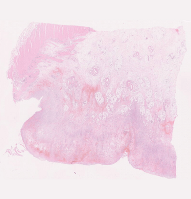
28. A 38-year-old male had multiple myxoid nodules on the peritoneum of the omentum. The nodules were 0.5 to 3 cm in size. One year after diagnosis, the patient seems to be healthy.
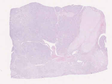
29. A 57-year-old male with a 7 cm deep-seated mass in the left thigh.

30. A 20-year-old male with lung metastasis. There was history of a bone tumor in the humerus two years ago.
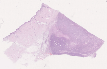
31. A 76-year-old male patient developed an indurated, dermo-subcutaneously located neoplasm on his back that was completely excised.
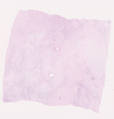
32. A 56-year-old male presented with a conglomerate of four dermal to subcutaneous tumor nodules on his left shin. The tumors elevated the overlying skin; their overall size was 3x2.5x2.5 cm. Enlarged lymph nodes in the left inguinal area sized 4.7x4.4x3 cm were detected at presentation and were later shown to represent metastatic disease (slides submitted to the seminar come from the metastasis).
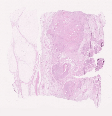
33. This 50-year-old male patient presented with a soft tissue mass in the right lateral hip. He had a past history of an injury to his hip 18 months previously that resulted in a golf ball-sized bruise and grew to be grapefruit in size at presentation. MRI of right hip in March 2016 revealed a well circumscribed 8.5x9 cm soft tissue subcutaneous mass of the right lateral hip without invasion of the gluteal muscles. The PET/CT scan showed no evidence of metastasis.
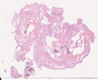
34. A 17-year-old male was seen for insidious onset of buttock pain. Imaging findings showed a large cystic tumor in his left sacrum involving S1–S3. The lesion was locally destructive and showed fluid levels suggestive of aneurysmal bone cyst (ABC). It was first attempted to control the tumor using radiofrequency ablation, which failed, and the patient was scheduled for surgery. A preoperative core biopsy showed small fragments of bone matrix associated with numerous large, atypical epithelioid cells with slightly eccentric nuclei, basophilic cytoplasm, and small nucleoli. The biopsy was regarded as atypical and suspicious for malignancy, but was felt to be inconclusive. At surgery, the tumor surrounded the nerve roots on the left side of S1, S2, S3, and the cauda equina. Embolization for hemorrhage control was done prior to surgery, causing the lesion to expand into adjacent vertebral levels and poke out of the foramina in front and in back of the bone. The tumor was excised and the area was bone grafted with internal fixation.
35. A 42-year-old man with a lipomatous tumor in the pectoral area measuring 4.8x3.5x1.4 cm.
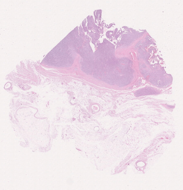
36. A 73-year-old male with an history of mucinous adenocarcinoma of the colon in 2013. The patient presented with an inguinal mass two years later, which was resected in another institution and relapsed one year later. No follow-up was available.
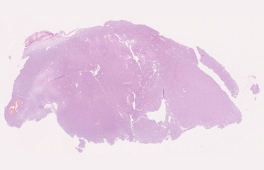
37. A three-year-old boy presented with a tumor on his upper lip that eventually invaded the orbit.
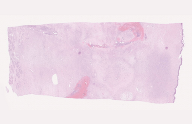
38. A 70-year-old man with a history of treated chronic myeloid leukemia was seen for development of left axillary soft tissue mass. A core needle biopsy was performed for the suspicion of myelosarcoma. The findings showed a high-grade-appearing sarcomatous neoplasm interpreted as a high-grade malignant spindle cell tumor consistent with malignant peripheral nerve sheath tumor. A wide excision of the lesion was performed. The specimen consisted of an ellipse of skin with attached underlying portion of the latissimus muscle and multiple small lymph nodes up to 2 cm in greatest diameter in the underlying fat. The tumor measured 6x5x4 cm and showed a white, glistening, homogeneous cut surface; the tumor reached up to the lateral and deep margins.

39. A 40-year-old woman with a 3 cm superficially located mass involving the vulvovaginal soft tissues.
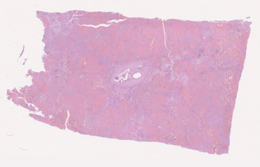
40. Patient is a 27-year-old female who presented with incidental splenomegaly comprising three splenic masses, characterized on MRI to be likely littoral cell angiomas or SANT, less likely a malignant process. These masses of the spleen were incidental and found when the patient had a CT to evaluate ovarian cysts. She reports no changes in her symptoms, still with a vague pressure in her abdomen two to three times per week. Otherwise, she feels well. No fatigue, weight loss, fevers, chills, or night sweats. Has poor appetite over the past several years with early satiety, but stable weight.
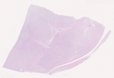
41. A 63-year-old woman with a 9 cm deep-seated mass involving the right thigh.
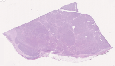
42. A 75-year-old woman with no significant past medical history was seen for shortness of breath and chest pain. CT scans revealed a large anterior mediastinal mass displacing the large vessels and compressing the innominate vein. A transsternal resection of the mass was undertaken, including an attached wedge resection of left lung, four mediastinal lymph nodes, and a portion of the innominate vein. The resected specimen contained a firm tumor mass, 7.5x6.7x5 cm, surrounded by adipose tissue. Cut section showed a fleshy, lobulated mass with areas of hemorrhage and cystic degeneration. There was a partial fibrous capsule surrounding the lesion, but focal infiltration of fat by tumor could be seen grossly.
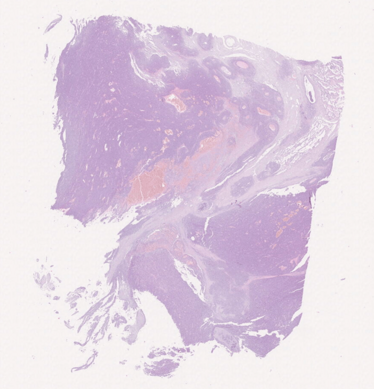
43. A 65-year-old woman with significant smoking history (40 py) presented with a 7.5 cm lung mass in the left lower lobe. She underwent left lower lobectomy and systemic lymph node dissection, followed by adjuvant radiotherapy. She presented 23 months later with an extensively invasive renal pelvis mass extending into the inferior vena cava, for which she received a radical nephrectomy with lymph node dissection. Thereafter, multiple metastases were detected by imaging in the liver, peritoneum, and in the inguinal, iliac, inter-aortocaval, para-aortic, and mesenteric lymph nodes. Under palliative treatment, the patient died of progressive disease 34 months from initial diagnosis.

44. A 54-year-old woman presented with a two-week history of epigastric pain. She was status post-cholecystectomy, appendectomy, and hysterectomy, and had a strong family history of malignancies. Her mother had colon cancer and uterine adenosarcoma, her father had colon and prostate carcinomas, her sister had basal cell carcinoma, two maternal aunts had gynecological cancers, one maternal uncle had gastric cancer, and her maternal grandmother had ovarian carcinoma. The patient underwent imaging studies and a 9 cm tumor was found in the upper abdomen (arising in the gastrohepatic ligament and extending to the abdominal wall and gastric serosa). A biopsy was obtained and the patient was treated with three cycles of neoadjuvant chemotherapy followed by surgery. Residual disease was confined to area mentioned above.
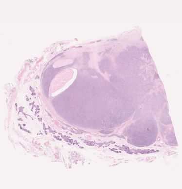
45. A 54-year-old man presented with symptoms of cough, chest pain, and dyspnea. Diagnostic imaging shows the presence of a large anterior mediastinal mass.
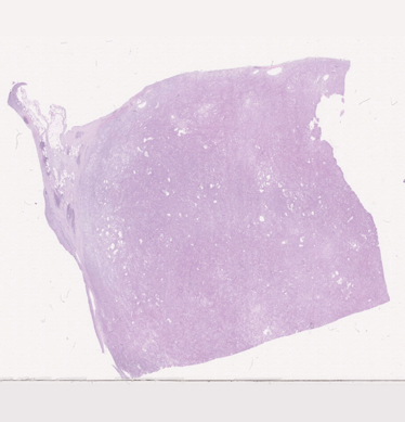
46. A 76-year-old man was diagnosed with a paraaortic intrathoracic mass initially thought to represent lung cancer. Pleural effusion cytology failed to demonstrate any malignant cells. Surgical resection was performed. Fifteen months later, he developed multifocal chest wall recurrences, for which he received palliative chemotherapy following subtotal resection.
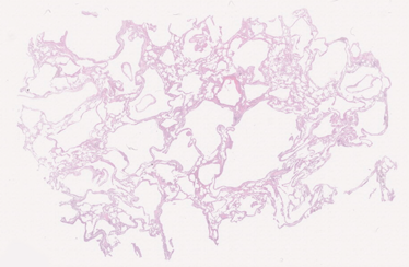
47. The patient was a 36-year-old woman who presented with progressive shortness of breath and pneumothorax. Chest X-ray showed some hyperinflation and mild reticulation suspicious for interstitial lung disease. Chest CT showed mild interlobular septal thickening and multiple thin-walled cysts of variable size distributed throughout the parenchyma of both lungs. She underwent a wedge biopsy of the left lung. The condition worsened over a two-year period and she underwent a left single lung transplant. She did well for approximately two years, but developed right lung herniation that required pneumonectomy. She developed postoperative complications and died with bronchopneumonia, chronic rejection of the left lung allograft, chronic renal insufficiency, and disseminated intravascular coagulation.
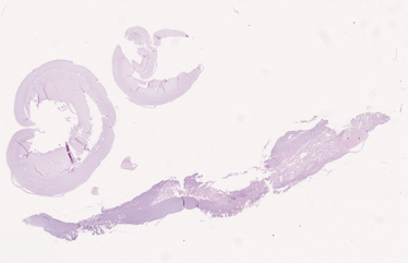
48. Adult male with lung “tumor.” Patient resides in Botswana.
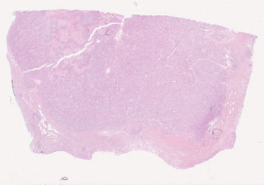
49. The patient is a 51-year-old woman, nonsmoker, with a past medical history of papillary thyroid carcinoma that was treated with partial thyroidectomy three years ago. She developed right hip pain. A chest X-ray performed during her workup showed an ill-defined density in the retrocardiac left lower lung zone. Chest CT showed a 3 cm spiculated mass suspicious for malignancy. PET-CT showed that the mass had an SUV of 21.7. She underwent mediastinoscopy and wedge biopsy of the mass. Frozen section showed an invasive adenocarcinoma. She underwent a left lower lobe completion lobectomy under the same anesthesia. Segmental and hilar lymph nodes showed metastatic tumor, so the lesion was staged as pT1b, pN1, pathologic stage IIA. The patient was subsequently treated with four cycles of cisplatin + pemetrexed chemotherapy. Postoperative radiation therapy was not recommended. The possibility of treatment with an ALK inhibitor was considered, but no clinical trial was available at the time of diagnosis.
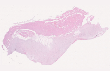
50. A 63-year-old man presented with shortness of breath, loss weight, chest pain, cough, and general malaise. Diagnostic imaging revealed the presence of thickening and nodularity of the right pleura.
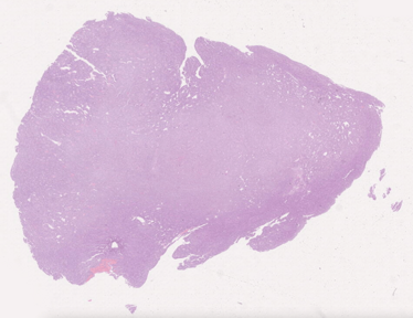
51. A 58-year-old woman presented with abdominal discomfort, bloating, and history of a 70-pound weight loss over the past year. A severe episode of abdominal pain led her to the emergency room at an outside community hospital. CT scan showed a 20 cm pelvic mass thought to be ovarian extending into abdomen, a 3.5 cm right hepatic lobe/diaphragm mass, moderate ascites, normal pancreas, and normal chest. The diaphragmatic mass was thought to represent a serosal implant. Serum levels for CA125 were 293.9, CEA 109 (normal <5), HE4 79 (normal <140), and CA19-9 14.7 (normal). Bilateral salpingo-oophorectomy, appendectomy, and resection of the diaphragmatic mass were performed. The patient is alive with no evidence of disease four years later.
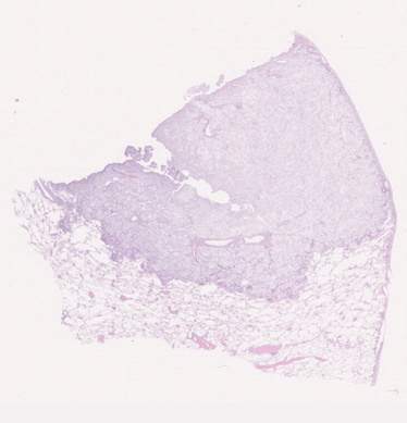
52. A 63-year-old woman was found during a routine chest film to have a mass in the right upper lobe. The mass appeared to be peripheral and subpleural. The patient had no history of malignancy anywhere else. Right upper lobectomy was performed, showing a 2.5 cm intrapulmonary mass.
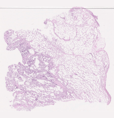
53. A 73-year-old female presented with an incidental 1.4 cm spiculated lung nodule found during a workup for longstanding back pain. She also complained of a new, non-productive cough for two months.
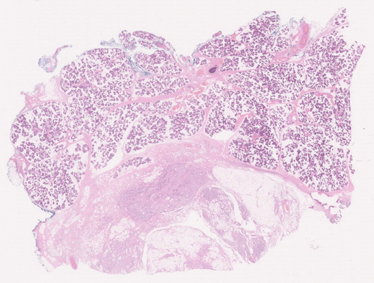
54. A 79-year-old man with hypertension and hyperlipidemia had noticed a painless, enlarging mass over the right parotid region for the past two weeks. On clinical examination, a 2.5 cm soft, well-delineated mass in the parotid region was noted. A FNA biopsy was performed followed by a CT scan that showed a heterogeneously enhancing, fairly well-circumscribed mass in the superficial lobe of the right parotid gland, measuring 2.5 cm. A superficial parotidectomy was performed. Grossly, the tumor appeared as a well-circumscribed, yellowish, soft mass measuring 2 cm in maximum dimension.
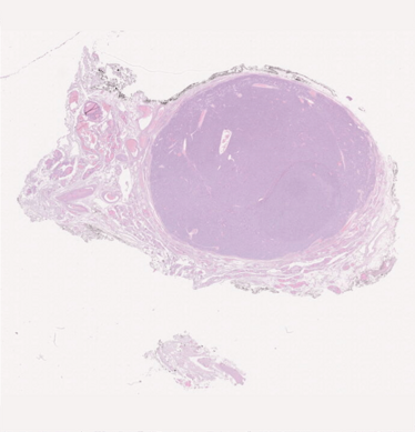
55. This 54-year-old female came for a consultation about treatment of her large, symptomatic right thyroid nodule, with FNA cytology showing a follicular neoplasm with positive NRAS mutation. Thyroseq suggested an 80 percent risk of associated thyroid malignancy.
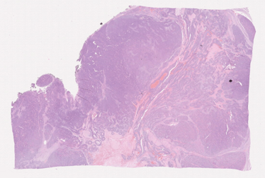
56. A 67-year-old man submitted to left lobo-isthmectomy for a 4.4 cm rapidly growing, locally invasive thyroid tumor.
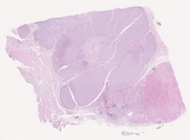
57. A 42-year-old female music teacher presented to her primary care provider after experiencing progressive throat tightness over the preceding year. She also noticed an anterior neck mass and bilateral cervical lymph nodes that had been gradually enlarging. The patient had been an otherwise healthy nonsmoker and social drinker. Family history included benign thyroid nodules in her mother and an unknown type of skin cancer in her father. Cervical sonography and computer tomography both revealed a 5.4x5.1x4.3 cm mass replacing the entire right thyroid lobe with extension into the isthmus. Also noted was bilateral central neck adenopathy and right lateral neck adenopathy. Fine needle aspiration was positive for papillary thyroid carcinoma. The patient consented to total thyroidectomy with bilateral central neck dissection and right lateral neck dissection.
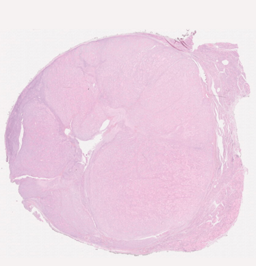
58. A 48-year-old woman submitted to total thyroidectomy (20 g). The right lobe was almost totally occupied by a well-circumscribed 3.6 cm tumor.
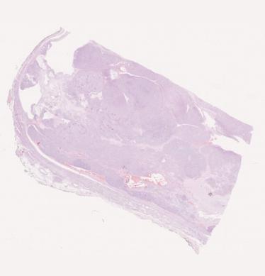
59. A 75-year-old woman with slowly enlarging thyroid and a large goiter on the right side, followed for several years. She also had HPT with a calcium of 10.9 and PTH of 93, with a possible parathyroid adenoma adjacent to the left, smaller side of the thyroid. CT scan and ultrasound showed a right lobe mass of 9 cm going from the parotid to just below the clavicle. She had several small nodules in the left lobe as well. FNA was performed on several occasions with benign results.
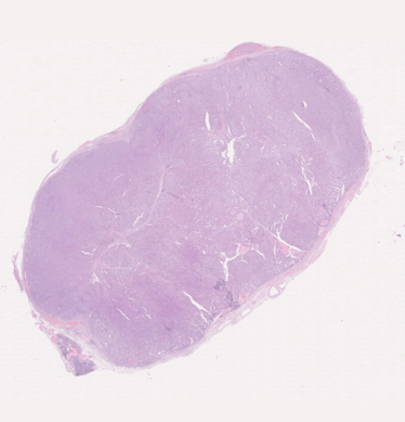
60. An 82-year-old female with a solid nodule (EU-TIRADS-4) in the right lobe of thyroid and multiple right cervical lymph nodes, the largest measuring 5 cm, detected on routine surveillance after a cardiac surgery for symptomatic severe aortic stenosis. Patient was submitted to total thyroidectomy with lymph node dissection.
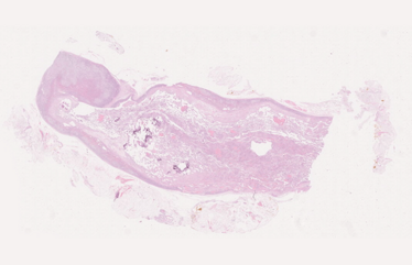
61. A 36-year-old woman presented with left lower flank point tenderness without a prior history of trauma to the area. In February 2017, an incidental 4 cm left adrenal mass was noted by CT scan. In March 2018, the mass measured 4.6 cm and was described as a complex cyst. A year later, in January 2019, the mass was 4.9 cm in maximum dimension. Serum levels of metanephrines, aldosterone, renin, and 24-hour urinary cortisol were all within normal limits. Most recent CT scan showed peripheral calcifications and internal layering with fluid-fluid levels. Although she was largely asymptomatic, the patient agreed to a robotic adrenalectomy.
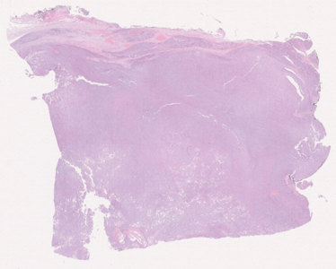
62. An 89-year-old woman presented with a left parotid gland mass that she defines was present for “a long time, but had increased in size in recent months.” She underwent surgery (superficial parotidectomy) for an 8 cm tumor that was poorly-circumscribed, elastic, and had a whitish cut surface. She was treated with postoperative radiotherapy and refused any further treatment. She is currently well, without recurrence (refusing treatment, but not follow-up consultations).
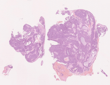
63. A 64-year-old female had a neoplasm that obliterated both nasal regions. The tumor was excised in several pieces. Originally classified as membranous variety of basal cell carcinoma, after HPV genotyping that revealed presence of HPV 56, the tumor was reclassified as HPV-related multiphenotypic sinonasal adenoid-cystic-like carcinoma.
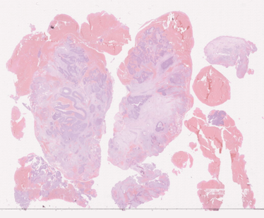
64. The patient was a 23-year-old woman with a few months' history of increasing left-sided nasal obstruction. On clinical examination, a large fleshy tumor occupying the whole (left) nasal cavity was seen. The tumor did not involve the right nasal cavity. On MRI, a tumor in the left nasal cavity was confirmed, extending from the anterior nasal cavity into the nasopharynx and from the anterior skull base to the inferior aspect of the inferior turbinate. Tumor extended into the left maxillary sinus and bulged into the orbit. The lesion obstructed the sphenoethmoidal recess and the ostiomeatal unit, resulting in fluid retention and mucosal thickening in the left frontal, ethmoid, and sphenoid sinuses. The overall radiological impression was that of a benign or low-grade malignant tumor. The possibility of an inverted papilloma was raised. The patient underwent a biopsy under general anaesthesia. After resection of the tumor, the patient underwent six weeks of chemo-radiation. There has been no evidence of local recurrence or metastatic disease with a follow-up of 4.5 years.
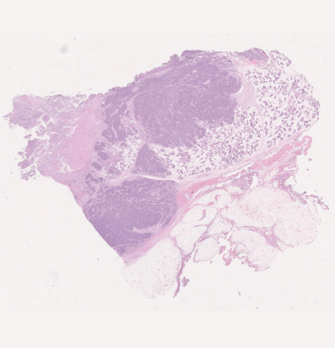
65. A 50-year-old woman presented to ENT clinic with a 3 cm firm, non-tender, rapidly enlarging right parotid mass that appeared FOUR weeks earlier. She now has lip weakness. Fine needle aspiration biopsy was interpreted as suspicious for lymphoma. CT of the neck revealed a 3.6 cm mass of the right parotid. She denies fever/chills and has no issues with swallowing. She has a history of invasive breast cancer treated with CRT 12 years earlier and no recurrence. A right parotid debulking was performed.
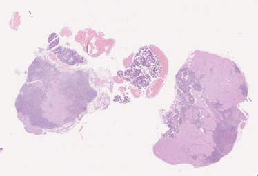
66. AN 82-year-old gentleman was referred because of a parotid gland nodule, 3 cm in largest dimension. He had enlarged lymph nodes at levels I and II. The homolateral lacrimal gland was also enlarged. He underwent surgery (the slides are from one of the lymph nodes). The patient died of unrelated causes in 2021.
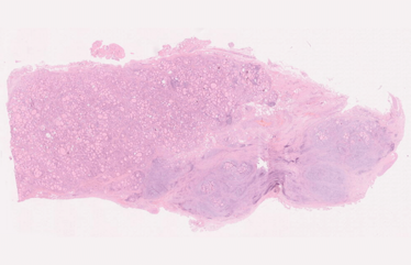
67. A 50-year-old male had a slightly enlarged thyroid with focally macroscopically fibrous white nodules.
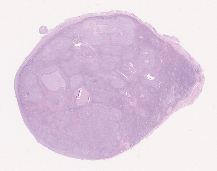
68. A 40-year-old man was referenced with cervical (levels I and II) lumps. He was otherwise healthy. From the history, no symptoms were reported. He was a smoker (~25 cigarettes a day) and had a brother undergoing therapy for Hodgkin lymphoma. He is currently well without recurrence of the neoplastic disease. The slide is from a level II lymph node.
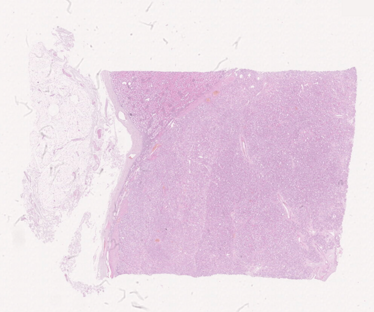
69. A 48-year-old female with a solitary renal tumor, size 2.5x3x2 cm, dark tan to brown in color, solid. No necrosis or hemorrhage.
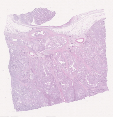
70. A 67-year-old female with a renal tumor located in the central part of the kidney. Diameter was 6 cm; tumor infiltrated renal sinus.

71. A 48-year-old female with hypertension and previous hysterectomy (leiomyomas) presented with 2–3 months' unintentional loss of weight, loss of appetite, and upper abdominal–left hypochondrium discomfort. She also had fever and anemia. She underwent a CT scan of the abdomen that revealed a 14.9x10.5x9.2 cm heterogeneous left renal mass that was inseparable from the left psoas, para-renal fascias, pancreatic tail, spleen, left ovarian vessels, and adjacent small and large bowel loops. There were nodular left perinephric deposits and a tumor thrombus left renal vein. Surgery (nephrectomy + extended anterior resection) was performed and the intraoperative findings were those of a large renal tumour involving the mesentery of the descending colon with tumor rupture. Four months after the surgery, a bone scan showed multiple bone metastases and she developed lung metastasis and non-ischemic cardiomyopathy. The patient received palliative chemotherapy.
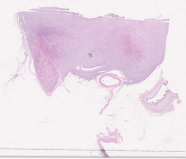
72. A 63-year-old female underwent a CT scan of the abdomen and pelvis for previous endometrial carcinoma and was incidentally found to have a 2.7 cm mass in the anterior interpolar region of the left kidney that was not appreciated during the previous year. The patient and urologist decided to perform a nephrectomy for definitive removal of the tumor.
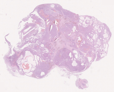
73. A 67-year-old male with tumor replacing majority of the kidney parenchyma. Metastases in regional lymph nodes and in adrenal gland were documented. Family anamnesis was unknown.
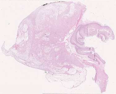
74. A 69-year-old female presented with “enlargement of the belly.” A CT scan showed a septated cystic mass that was ultimately excised and found to be a seromucinous cystadenofibroma of the left ovary. During the procedure, an incidental mass was seen in the wall of the distal small bowel. Frozen sections showed a spindle cell proliferation. Given a clinical concern for gastrointestinal stromal tumor, the remainder of the lesion was resected.
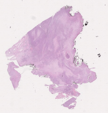
75. A 42-year-old male with supraorbital paresthesia and diplopia in 2007. CT scan revealed an intraorbital mass. This tumor was resected in another institution and relapsed in 2011, 2013, and 2018 after radiation therapy. The patient died after one year with diffuse metastasis.
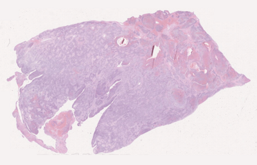
76. A 17-year-old woman with a history of multinodular goiter presented in 2011 with acute onset of heavy vaginal bleeding and was found to have an 8.5 cm hemorrhagic mass protruding from the vaginal introitus. The polypoid mass originated from the cervix and was surgically excised. Six years later, the patient presented with a cervical polyp and a 7.5 cm left adnexal mass. Representative blocks from the original cervical mass (2011) were submitted for this seminar.
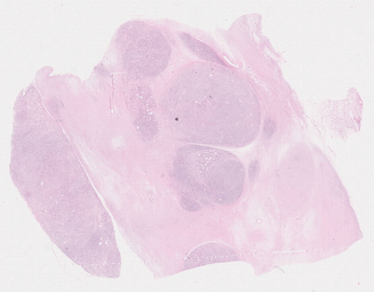
77. A 61-year-old female patient developed a 9x7x5 cm neoplasm at the vulva that has been marginally excised.
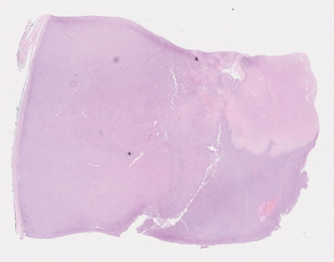
78. A 28-year-old male with abdominal pain was found to have retroperitoneal mass. Excision of the mass was performed. The patient was treated with three different lines of chemotherapy; however, he never achieved complete remission and died of disease approximately two years after diagnosis.

79. A 71-year-old male with tumor in the left kidney 6.5 cm in size. The patient lacked family history of sickle cell trait or other hemoglobinopathies. Radical nephrectomy with regional lymphadenectomy was performed. Multiple metastatic sites were apparent at bone CT scan. The patient died three months later.


