Guillermo Tearney

Professor of pathology at Harvard Medical School and an affiliated faculty member of the Harvard-Massachusetts Institute of Technology (MIT) division of health sciences technology, Guillermo received his MD from Harvard and his PhD in electrical engineering and computer science from MIT. His research is focused on developing and clinically validating noninvasive, high-resolution optical imaging methods for disease diagnosis. “I think pathologists are the most qualified people to be diagnosing in vivo microscopy, and have a major role to play in its adoption.”
Guillermo (Gary) Tearney is a Mike and Sue Hazard Family MGH Research Scholar, Professor of Pathology at Harvard Medical School, an Affiliated Faculty member of the Harvard-MIT Division of Health Sciences and Technology (HST), and maintains his lab at the Wellman Center for Photomedicine at the Massachusetts General Hospital. Guillermo received his MD magna cum laude from Harvard Medical School and his PhD in Electrical Engineering and Computer Science from MIT.
His research interests focus on the development and clinical validation of non-invasive, high-resolution optical imaging methods for disease diagnosis. Guillermo’s lab was the first to perform human imaging in the coronary arteries and gastrointestinal tract in vivo with Optical Coherence Tomography (OCT), which provides cross-sectional images of tissue architectural microstructure at a resolution of 10 µm. He has also conducted many of the seminal studies validating OCT and is considered an expert on OCT image interpretation. Recently, his lab has invented a next generation OCT technology, termed µOCT, which has a resolution of 1 µm and is capable of imaging cells and subcellular structures in the coronary wall.
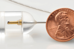
Other technologies he has developed include a confocal endomicroscope capable of imaging the entire esophagus, the world’s smallest endoscope, a microscope capable of imaging at the nanoscale, and novel spectroscopy and multimodality imaging techniques. He has an active program in Raman spectroscopy and has conducted the first intracoronary Raman in vivo. He is co-editor of The Handbook of Optical Coherence Tomography and has written over 200 peer-reviewed publications, including papers that have been highlighted on the covers of Science, Nature Medicine, Circulation, Gastroenterology, and Journal of American College of Cardiology.
With over 400 patent applications (~80 granted) and licenses on more than 200 patents/applications resulting in commercial medical devices, Guillermo has also been a principal investigator on over 40 grants. His work extends beyond the lab, with many of his technologies being produced commercially.
He is the chair of the Research Advisory Committee for the Institute for Aging Research, a member of the Scientific Advisory Board for Massachusetts Life Sciences Center and the Clinical Advisory Board for NinePoint Medical, and he has founded the International Working Group on Intravascular OCT Standardization and Validation, a group that is dedicated to establishing standards to ensure the widespread adoption of this imaging technology. “I think pathologists are the most qualified people to be diagnosing in vivo microscopy, and have a major role to play in its adoption.”


