Using Whole Slide Imaging for Primary Diagnosis: An implementation journey and future directions
In this webinar Dr. Parwani will discuss how he and his team are continuing to work towards a “slideless” workflow.
sponsored by Philips

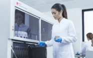
In the 16th century the microscope was first invented; this revolutionized the diagnosis of disease. Since then a lot has changed, most notably increasing use of technology which has transformed our lives and in particular within healthcare. Today pathology services are under mounting pressure to provide efficient and high-quality diagnoses with diagnostic workloads increasing in volume and complexity. Leveraging innovative and intelligent digital solutions can help support pathologists in delivering quick and accurate diagnoses, while simultaneously lowering the cost of healthcare delivery through increased operational efficiencies.
Dr. Anil Parwani is a Professor of Pathology at The Ohio State University, where he has worked with a team of IT specialists, vendors and histology staff and pathologists to successfully implement high throughput whole slide imaging for primary diagnosis. In this webinar Dr. Parwani will discuss how he and his team are continuing to work towards a “slideless” workflow.
Learning objectives of webinar:
- Discover why The Ohio State University decided to implement digital pathology
- How the team at OSU selected the right system for their laboratory, how this was validated and the what was the business case behind going digital
- What are the advantages and limitations of now diagnosing digitally and future directions


