“ScanVan doesn’t start with technology. It starts with Katie.”
Todd R. Minnigh – Chief of Medical Imaging Innovations at Leidos – is the model of a proud family man as he shows me photos of his beloved niece, Katie, standing beside his daughter, Sarah, and her older sister, Delaney. It’s an ordinary snapshot. The kind that doesn’t hint at what’s coming.
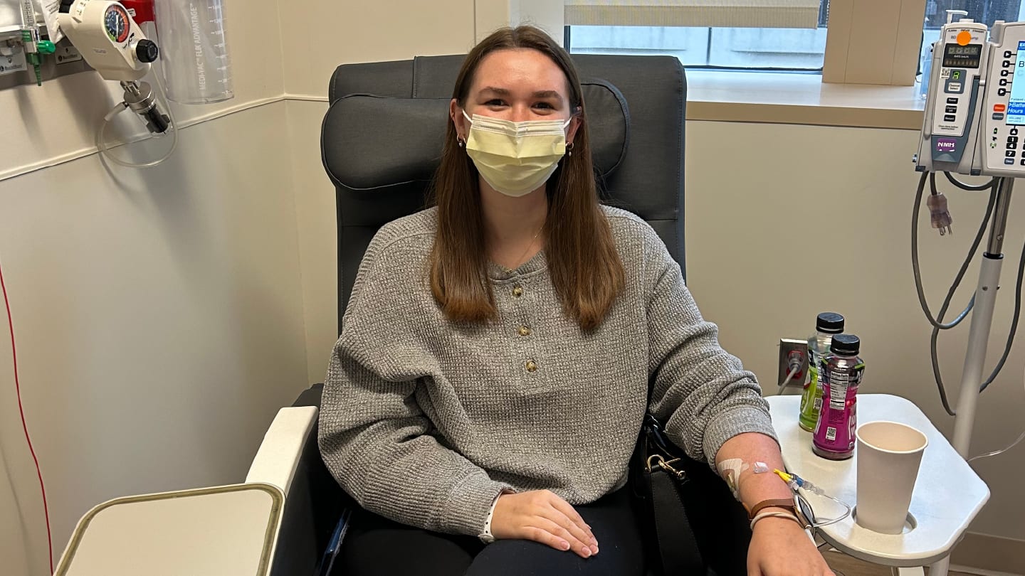
In October 2022, Katie noticed a lump in her abdomen. Her doctor suspected a hernia. Routine. Fixable. She scheduled surgery at a small hospital outside Chicago. But in the operating room, the story changed.
“The lump was in fact some kind of sarcoma,” Minnigh says. “They weren’t exactly sure what kind.”
The 110-day delay
What followed is familiar to anyone working in rare disease diagnostics: uncertainty, escalation, and distance. Katie was referred to another hospital in Chicago. There, clinicians began to narrow the possibilities.
“The interim conclusion was, we think this is something called alveolar soft part sarcoma,” Minnigh explains. ASPS is astonishingly uncommon. Most pathologists will encounter it rarely, if at all. For Katie’s family, that rarity quickly translated into geography. Their research pointed them to a single center with deep experience: Dana-Farber Cancer Institute in Boston.
“So, off to Boston,” Minnigh says. Tissue blocks, slides, and data were physically shipped. Then, eventually, Katie herself followed. In Boston, specialists reopened the case, reviewed the material, and confirmed the diagnosis: ASPS.
Meanwhile, the initial surgery triggered Katie's tumor to metastasize, leading to follow-up surgery and the loss of a lot of abdominal tissue. “From the time that she has the operation in the suburbs of Chicago until she’s being treated for what she has in Boston,” Minnigh says, pausing, “110 days goes by.”
He lets the number sit for a moment. “Did it need to take 110 days? Well… probably not.”
Access to expertise
The reasons are not mysterious. They are structural. Physical specimens move slowly. Expertise is centralized. Scheduling takes time. And in rare cancers, diagnostic certainty often depends on a few specialists, concentrated in a handful of institutions.
“Imagine if that doctor in Chicago could have taken the frozen section and put the image up on a database that said, ‘Hey, it’s probably this. And if it is, here’s the safest protocol to follow.' The decision time could have been reduced from 110 days to 20 minutes.”
The problem is not a lack of expertise. It’s access to it, at the moment it matters most. This is where Minnigh sees the role of artificial intelligence. Not as a replacement for pathologists, but as a way to extend their reach. To make expertise available in real time, wherever the patient happens to be.
Making smart oncologists smarter
“The spirit of this is, what if we could make smart people smarter?” he says. “What if we could give physicians AI to assist with unfamiliar cases?”
But building that kind of system comes with its own challenge: “To create AI, you need thousands of slides. You need to curate them and annotate them, and you need them in high resolution.”
Those slides exist. In fact, they exist in staggering numbers. In other words, the raw material for smarter, faster diagnosis already exists. It’s just not accessible in the way modern medicine demands.
For Minnigh, that gap between potential and reality became the next problem to solve.
The rarity problem
In the US, The National Cancer Institute defines cancer that occurs less than 40,000 times a year as rare. If you spread those cases over all the hospitals in the US, it equates to zero to a handful per year at each site.
“Even for the most common of these rare cancers, if a hospital keeps those slides for 10 years, they’re only going to have, say, 75 cases at any one given hospital,” Minnigh explains. “It makes the disease really difficult to study.”
However, there’s a pattern to where rare cancer cases end up. Patients, especially younger ones, don’t stay local for long, but seek out specialist experience at cancer centers and university hospitals. Over time, that movement reshapes the data landscape. Tissue flows toward expertise. Instead of covering 5,000 fragmented sites, the real number is closer to 400 institutions where meaningful volumes accumulate.
That changes the picture. For some of the more common rare cancers, there’s enough material to begin building datasets, spotting patterns, and training algorithms. That's what Minnigh wants to access. “This project is to aggregate the data from many places,” he says.
It’s a simple idea, but a difficult one in practice. Data lives in different systems, different formats, and often still on glass. Moving it is slow, expensive, and sometimes impossible.
Unless, Minnigh began to wonder, you stop trying to move the data – and start moving the lab instead.
From archive to discovery
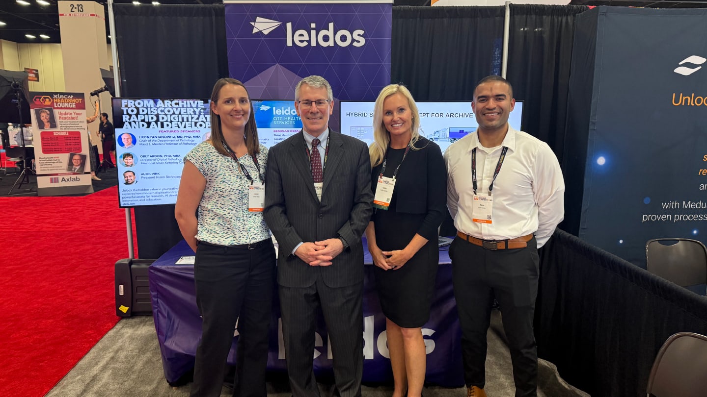
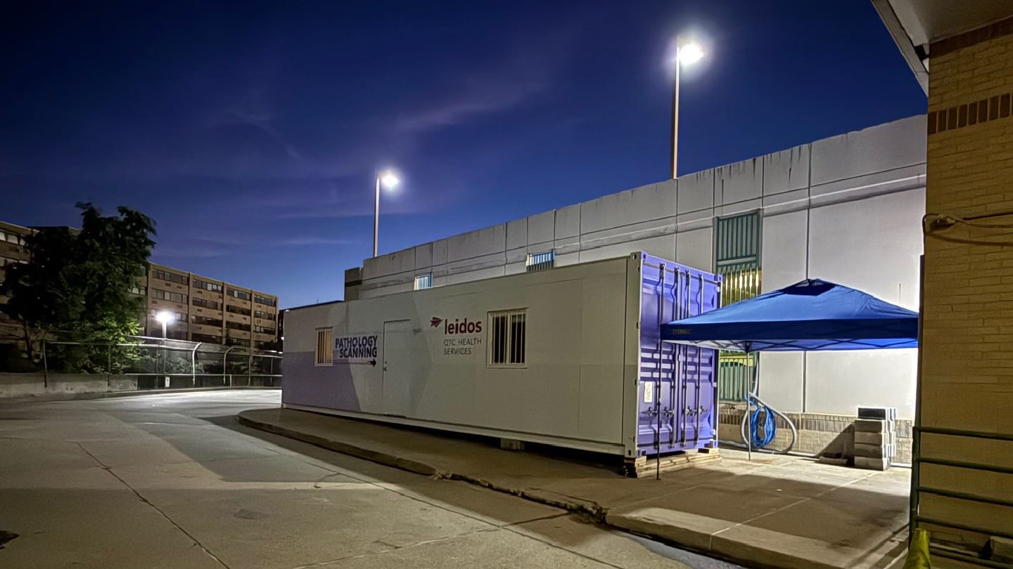
“ScanVan is a mobile digital laboratory in a shipping container,” Minnigh explains, with pride. “The eight slide-scanners on board have the capacity to digitize around 1.1 million slides a year.”
The numbers are important. Rare cancers, while small in isolated incidence, collectively account for around 25 percent of all cases. If ScanVan visits a hospital with an archive of 100,000 cancer slides, roughly 25,000 will contain rare cancer samples.
“In less than a month, we can scan all the rare cancers, shut it all down, pack up, and move to the next place,” says Minnigh. “We're just finishing up at the University of Pittsburgh Medical Center, where the Computational Pathology and AI Center of Excellence (CPACE) helped us automate and prove out the ScanVan prototype. Next, we're moving to the Veteran's Affairs hospitals, starting at the Tampa VA Medical Center.”
And so the data aggregates. For each cancer type, a critical mass of data accrues for research, education, and AI modelling. Its reach extends outside the pathology lab. “The pharma companies believe they can use it for biomarker discovery,” he says.
This database approach could spark a paradigm shift in rare cancer research. “Say I want to study squamous cell carcinoma of the larynx and trachea,” Minnigh explains. “I go from one cancer center to another, seeing if I can assemble a couple of hundred physical samples. That is a heavy lift to address just one very rare disease.” Meanwhile, ScanVan's parallel processing approach is building a digital archive of thousands of samples of each rare cancer.
But before the initiative could get off the ground, there were some challenges to navigate.
Filling in the potholes
The beauty of such a single-minded scanning facility is that it can be built around efficiency and trouble-shooting. Its secret? Pre-sorting the slides.
“We put most of them through the Leica scanners, which are small and fast,” says Minnigh. “If the slide is dirty, the cover slip is cracked, or has artifacts, we run it through Pramana machines. While they are larger and more expensive, they are also more fault-tolerant.”
Quality control of scanned images also required a considered approach – one operator can't do all the QC and maintain the scanning rate. That's where technology comes in. “Aira Matrix has been kind enough to let us test its QC software,” shares Minnigh.
However, even the best quality data are meaningless if they can't be retrieved. And when retrieval relies on barcoding systems that vary not only between institutions, but often within them, it's far from straightforward. “Creating a system that allows slide images to be found later on is one of the major hurdles we've faced,” he reflects.
After scanning comes data transfer back to the hospital system – a process fraught with red tape: “IT people don't want you to show up with a bunch of non-approved equipment and plug into their network.” Fortunately, multi-terabyte hard drives solved that problem, allowing hospitals to download batches of data after thorough security scanning.
One by one, Minnigh and his team smashed the barriers, refined the process, and got the van on road. After all, they had a population health crisis to address.
A question of statistics
Now let's return to Katie. How did her story unfold?
“Katie's still being treated for the metastasized tumors.” Minnigh reflects. “That's the reality of the consequences of diagnostic delays. That's why we are doing this.”
“But she's doing great... now in her late 20s, thriving in a great job in downtown Chicago, winning awards for writing about her experiences...” He's a proud and relieved uncle.
“Were it not for Katie, I would probably be retired,” he laughs. “So I have this project, and it's so close to what's happening with her, I feel compelled to get it off the ground.”
And if his vision spreads, ScanVan's legacy might be to prevent other people's loved ones becoming just another cancer statistic.
Newsletters
Receive the latest pathologist news, personalities, education, and career development – weekly to your inbox.