Spitz nevi were first recognized in the early 20th century, following Sophie Spitz’s 1948 description of the so-called “melanoma of childhood.” In her landmark paper, she noted that these lesions could appear worrisome under the microscope but typically followed a benign course. Over the following decades, continued refinements in histopathologic criteria and, more recently, molecular discoveries have greatly advanced the classification and understanding of these lesions.
Spitz’s 1948 report introduced the term "juvenile melanoma" to describe a histologically alarming yet clinically benign lesion, sparking decades of discussion in dermatopathology. Later, the condition was named "Spitz nevus" in her honor, although distinguishing atypical variants from melanoma remained challenging.
This article examines the background of this pioneering female scientist and the remarkable impact of her controversial research.

Who was Sophie Spitz?
Sophie Spitz was born and educated in Nashville, Tennessee, USA. Her uncle, Herman Spitz, was a clinical pathologist, and it was through him that Spitz was first exposed to the medical world.
That early exposure likely inspired Spitz's decision to study medicine at university – a highly unusual move for a woman at that time. Undeterred by convention, she graduated from Vanderbilt University School of Medicine in 1932.
Pursuing a keen interest in pediatric pathology, Spitz completed a pathology residency at the New York Infirmary for Women and Children. She then stayed on in New York, accepting a position as assistant attending pathologist at the Memorial Sloan Kettering Cancer Center in 1941.
There, she met her husband, Arthur Allen, a fellow pathologist, and they married in 1942. With World War 2 waging, Spitz was called to serve her country at the Army Institute of Pathology. But she returned to Memorial Sloan-Kettering after the war to pursue her academic research.
Spitz was eventually promoted to Director of Pathology at the New York Infirmary, where she remained until her death in 1956.
Spitz's legacy
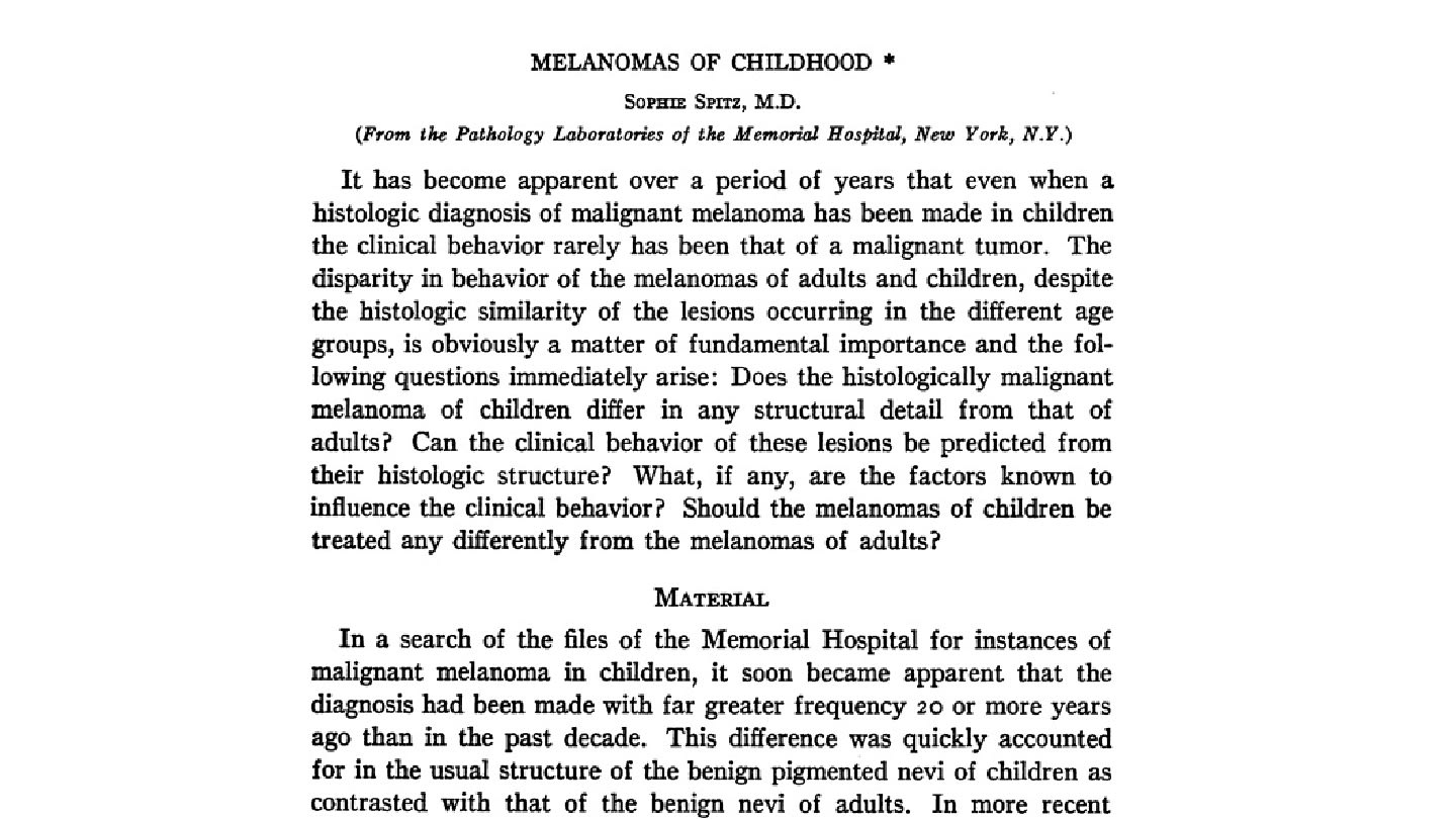
Spitz's seminal paper, Melanoma's of Childhood, was published in the American Journal of Pathology in 1948 (Figure 1). In it, she presented histologic evidence that tumors diagnosed as malignant melanoma were usually benign in children. Her findings supported a new classification of the condition in children: benign juvenile melanoma.
Research into the newly identified condition continued through the 1960s to 1980s. In 1967, the eponym “Spitz nevus” became established in Spitz's honor. However, while pathologists gradually refined the histologic criteria of the tumors, they struggled to separate atypical spitzoid lesions from true melanoma.
In 1979, Kamino and colleagues recognized the PAS-positive, diastase-resistant pink bodies often seen on H&E slides in Spitz nevi. They have been referred to as Kamino bodies ever since.
By the late 1990s, HRAS mutations and chromosomal 11p alterations were identified as the first molecular determinant of Spitz lesions, fundamentally differentiating Spitz nevi from other melanocytic tumors with spitzoid morphology. In 2009, the SPARK nevus variant, combining features of Spitz and Clark (dysplastic) nevi, was formally described.
In 2014, Wiesner and colleagues reported frequent kinase fusions involving ALK, NTRK1, and ROS1 in approximately 50 percent of Spitz neoplasms, offering diagnostic and potential therapeutic targets, and refining risk stratification.
More recently, in 2022, the concept of melanocytoma was solidified in the classification of intermediate-risk spitzoid tumors, aligning molecular biology with histology and clinical behavior, and evolving into the WHO classification of skin tumors, 5th edition (Figure 2).

A practice-changing discovery
Although Spitz's theory was initially challenged, her paper has since been cited more than 700 times and is widely recognized as having transformed pediatric melanoma diagnosis. As the medical world began to accept her research, the standard of practice for childhood melanocytic lesions, was finally changed ‒ sparing countless children from unnecessary and aggressive cancer treatment.
The history of the Spitz melanocytic neoplasm reflects a fundamental shift in dermatopathology, from reliance on morphology alone to a molecularly integrated framework for diagnosis and risk assessment. Ongoing advances continue to refine classification and management strategies, reflecting the ongoing evolution of pathology.
During an era when only six percent of physicians were women, Sophie Spitz broke barriers in pathology. She died at age 46 from metastatic colon cancer, leaving an enduring legacy that transformed dermatopathology.
Newsletters
Receive the latest pathologist news, personalities, education, and career development – weekly to your inbox.


