Has cancer always plagued the human race? That’s hard to answer with certainty, but researchers have discovered that our ancestors may have been fighting the disease much earlier in history than expected – over 4,000 years ago (1).
We connected with Tatiana Tondini, one of the lead researchers in the study, to learn more about this discovery.
Why did you choose to revisit analysis previously conducted on both skulls?
There are very few paleopathological records of cancer – only 270 confirmed cases globally – and these two skulls accessed through the Duckworth collection were known in relation to ancient cancer. Skull 236 was studied in 1963 and identified with nasopharyngeal carcinoma, but without access to modern tools available today, we theorized that a lot of crucial patient data could easily have been missed.
Alternatively, skull E270 had never been formally analyzed and had no published records – it had purely been used for teaching material showcasing cancer's effect on bones. Rumors suggested meningioma was present, but our research discarded this idea.
What challenges did you face?
Working with old and weakened skulls made this project quite difficult, especially handling the specimens for microscopic analysis. This is why we used a special soft support to prevent further damage during analysis.
The lack of records also brought about different challenges. We could contextualize each specimen, but understanding the archaeological context was another matter entirely. Additionally, only having access to the skulls of each individual meant we couldn’t work with a complete picture of the patients.
Of course, both skulls had several taphonomic damages (post-mortem breakages, often caused by environmental factors) – especially skull 236. Using a powerful microscope, we could characterize most lesions, and micro-CT scans allowed us to analyze the internal structure of more complex lesions and determine the cause.
Please tell us more about the use of micro-CT scanning and microscopic bone surface analysis
Skull E270 was analyzed with a Bruker Skyscan 1273 microCT scanner (165 kV, 128 μA, 71.274 μm), whilst Skull 236 was analyzed with a Nikon XTEK H 225 ST MicroCT scanner (130 kV, 100 μA, 81.020 μm). Micro-CT scanning has been particularly useful in providing us with a complete overview of the internal and external aspects of both skulls. Skull 236 presents 16 visible cancer lesions on the outer surface and this technology allowed us to identify 30 internal cancer lesions, as well as better characterizing the primary tumor on the palate.
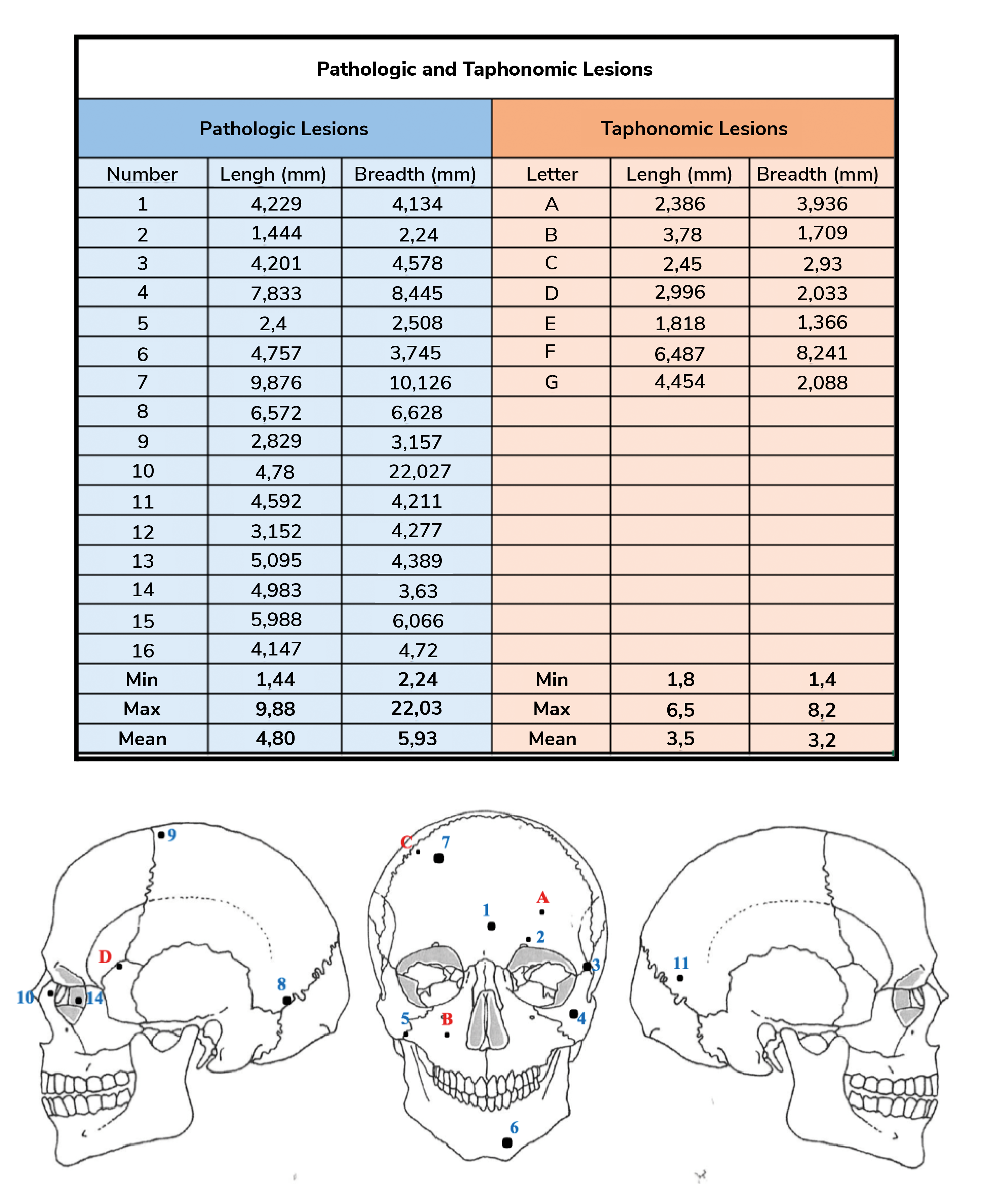
List of the measurements of length and breath of the pathological and taphonomic lesions on
skull 236 (along with the calculation of mix, max, and mean).
The Micro-CT scanning of skull E270 has also allowed for a more complete differential diagnosis and identified osteosarcoma as the most likely cancer affecting the patient. We could also see the severity of two traumatic lesions and the healing process.

Micro-CT images of skull 236 examined using DataViewer and Dragonfly. Reconstruction of
virtual images (coronal plane) that show the internal lesions indicated by the red arrows.
What were the key findings of your research?
The most remarkable finding was identifying cut marks near two secondary cancer lesions. These were confirmed to not be taphonomic (environmental) damage, leaving two explanations: i) the ancient Egyptians tried to surgically remove the secondary tumors in the patient's brain, which would make it the first recorded cancer surgical operation in history that we’re aware of, or ii) the ancient Egyptians left these traces when examining the body post-mortem, which hints at forensic autopsy and studying of cancer more than 4,000 years ago.
Another important finding resides in the successful treatment of the frontal bone lesion of skull E270. Our analysis showed this lesion was caused by a sharp object, like a sword or hatchet. Previous knowledge would lead us to believe that this patient couldn’t have survived. However, we can see clear signs of healing, which means that ancient Egyptians were able to treat severe cranial fractures.

Micro-CT images examined with DataViewer and Dragonfly used for the reconstruction of
virtual images (coronal plane): (a) Images showing the different angulations from which the skull was
analysed along with a virtual reconstruction showing the three lesions. Lesion I suggests the presence of
the Codman's angle; (b) Image shows the cancer destruction; (c) Codman's angle is visible; (d) Codman's
angle is visible as well; (e) Image shows the destruction of the cancer as well as the lesion 3 (depression
lesion); (f) Image shows the skull in cross section.
What’s next for the future of cancer and forensic research?
The next step of our research would involve molecular analysis of both skulls, such as ancient DNA, ancient proteins, and isotopes. However, this implies taking bone samples, which is very risky on ancient and fragile specimens. Since this research project utilized only non-destructive methods and still resulted in exciting discoveries, we hope to stimulate more research on cancer cases in antiquities using non-destructive methods.
Image Credits: Tatiana Tondini and colleagues, Institute for Archaeological Sciences, Eberhard Karls University of Tübingen, from their research on ancient cancer, 2024
Newsletters
Receive the latest pathologist news, personalities, education, and career development – weekly to your inbox.

References
- T Tondini et al., Front Med (2024). PMID: 38868751.


