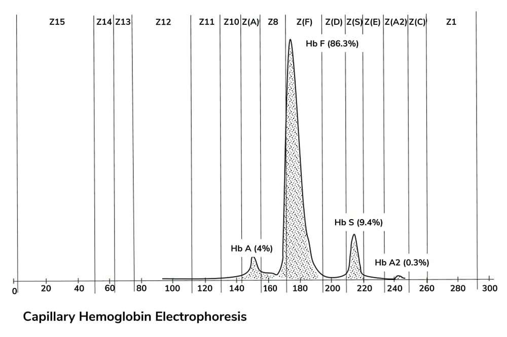
A two-day-old boy of Nigerian descent was tested for hemoglobin disorders. His father was known to have sickle cell trait. The patient’s sickle cell solubility test was positive. His red cell indices, serum ferritin, and hemoglobin electrophoresis histogram by capillary electrophoresis are shown below:


What is the best diagnosis for this newborn?
a. Sickle cell trait (Hb AS)
b. Sickle cell anemia (Hb SS)
c. Sickle beta-plus thalassemia (Hb Sβ+ thalassemia)
d. Sickle beta-zero thalassemia (Hb S/β0 thalassemia)
Click here to register your guess.
Do you have an interesting case that you would like us to feature? Email it to edit@thepathologist.com.
Answer to December's Case of the Month


C. Verrucous pseudonevoid melanoma
Histopathology revealed marked epidermal hyperplasia and papillomatosis overlying a broad, atypical melanocytic proliferation. Melanocytes were seen proliferating in a confluent fashion in both nests and solitary units at and above the dermo-epidermal junction. An uneven, sheet-like proliferation of small, uniform-appearing melanocytes was seen in the upper dermis. Melanocytes arranged in parallel cords – the so-called parallel thèque pattern (1) – with surrounding sclerosis were observed. Rare dermal mitoses were identified and Ki-67 stain (not shown) revealed a proliferation index of approximately 10–15 percent in the dermal melanocytes.
The overall presentation was most consistent with a verrucous pseudonevoid melanoma of the scalp. The Breslow depth was measured at 1 mm. The patient was treated with a wide local excision with 1 cm margins. The re-excision specimen showed focal residual melanoma in situ and clear margins. A sentinel lymph node biopsy was performed for additional prognostication and was found to be negative for metastatic disease, yielding a final stage of 1a.
Nevoid melanoma is a rare entity that presents significant diagnostic difficulty on both clinical and histopathological grounds (2,3,4,5). On physical examination, this tumor can be mistaken clinically for a verruca, benign melanocytic or epidermal nevus, or seborrheic keratosis. Two major architectural variants have been reported: verrucous subtype and a dome-shaped variant (resembling a Meissner or Spitz nevus). The verrucous subtype (as seen in this case) has the following features that may distinguish it from a papillomatous nevus: i) broad, exophytic growth pattern with verrucous epidermal hyperplasia, ii) continuous proliferation of melanocytes along the dermal-epidermal junction, iii) confluent sheets of small- to medium-sized melanocytes in the dermis without evidence of true maturation, and iv) low, but appreciable dermal mitotic activity.
Risk of mortality in nevoid melanoma is thought to be consistent with that of traditional melanomas of the same Breslow depth. Nevoid melanomas are often more advanced at the time of diagnosis given the propensity for initial clinical or histologic misdiagnosis. Heightened awareness of this entity is critical to promote earlier biopsy and avoidance of misdiagnosis (6).
Submitted by Rand Abou Shaar, Department of Pathology; Joseph McGoey, Department of Dermatology; and Ben J. Friedman, Department of Dermatology, Henry Ford Health System, Detroit, Michigan, USA.
References
- MH Idriss et al., J Am Acad Dermatol, 73, 836 (2015). PMID: 26299955.
- TY Wong et al., Hum Pathol, 26, 171 (1995). PMID: 7860047.
- C Schmoeckel et al., Arch Dermatol Res, 277, 362 (1985). PMID: 4026378.
- A Zembowicz et al., Am J Dermatopathol, 23, 167 (2001). PMID: 11391094.
- K Blessing et al., Histopathology, 23, 453 (1993). PMID: 8314219.
- R Cabrera, F Recule, Am J Clin Dermatol, 19(Suppl 1), 15 (2018). PMID: 30374898.


