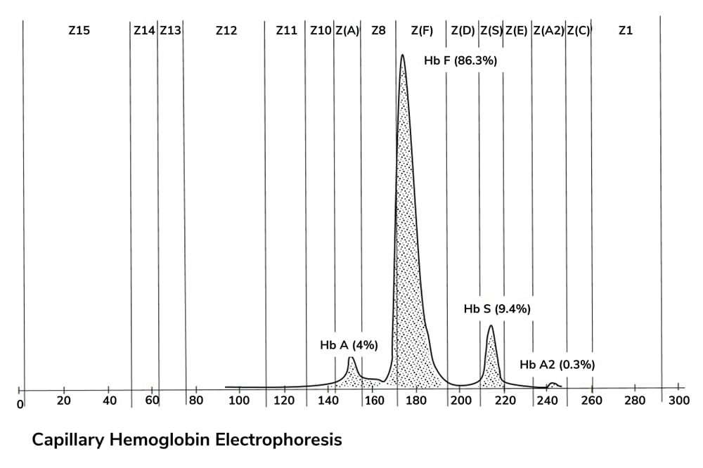
- Glycated hemoglobin (HbA1c) testing, the standard method of long-term glucose monitoring, can be inaccurate in patients with abnormal red blood cell turnover
- Glycated serum protein (GSP) levels are similarly reflective of average blood glucose levels and could provide a suitable additional measure
- GSP testing is an intermediate marker of glycemia, providing measurements for a two- to three-week period that bridges the information gap between short- and long-term monitoring
- Combining HbA1c and GSP testing offers improved diagnostic accuracy and reliability in the prediction of diabetic complications, especially in patients with conditions that affect red blood cell lifespan
Since blood glucose monitoring began in the 1960s, it’s been a key parameter for the control of acute diabetes. It has made daily glucose monitoring by patients possible, and, for longer-term control, glycated hemoglobin (HbA1c) assays have traditionally been the primary test used in clinical practice, offering average measurements over a two- to three-month period. But there is a gap which is, as yet, largely unplugged – the gap between short- and long-term testing. Given the exceptionally high prevalence of diabetes, and a projection that the disease will be the seventh leading cause of death worldwide by 2030 (1), it’s clear that meticulous blood glucose tracking and management – short-, medium-, and long-term – is of great, and growing, importance. Until recently, there was no reliable marker for medium-term monitoring. But now, a new marker, glycated serum protein (GSP), could be the answer. It could also provide a suitable alternative to HbA1c in patients with abnormal red blood cell turnover.

The standard measure for glucose control
Along with daily blood glucose measurements, HbA1c is commonly tested in patients with diabetes because it provides a reliable measure of glycemic control. Circulating blood glucose irreversibly attaches to hemoglobin A in red blood cells, making the hemoglobin molecules highly stable (Figure 1). As a result, HbA1c levels reflect a weighted average of circulating glucose levels over the two- to three-month lifespan of red blood cells, giving clinicians an important piece of information about their diabetes patients’ long-term blood glucose management. The HbA1c assay can also be used as a diagnostic test for diabetes, allowing doctors to identify at-risk individuals early and helping them make small lifestyle changes to reduce their chances of developing type 2 diabetes. All of these uses make HbA1c the standard in the diabetes world – for prevention, diagnosis and management of the disease.At the moment, HbA1c is primarily used to track long-term trends in blood glucose for diabetic patients; the amount of glycation present on the hemoglobin proteins is measured against the total amount of hemoglobin in the red blood cells. But a technique that relies on the lifespan of a red blood cell has its limitations. Critically, HbA1c testing is not suitable for patients with conditions that affect red blood cell turnover – hemoglobinopathies, thalassemias, chronic and end-stage kidney disease, and some forms of anemia (2). Factors such as age, race, pregnancy, and certain drug treatments can also skew the HbA1c measurement. In patients like these, an alternative monitoring method is needed.
Closing the gap
Like hemoglobin, serum proteins also undergo non-enzymatic and irreversible glycation. Albumin, also known as fructosamine, is the most abundant of the serum proteins, and glycated albumin (GA) accounts for 80 to 90 percent (3) of the GSP test readout. GSP is also strongly correlated with HbA1c and mean blood glucose in type 1 and type 2 diabetes (4,5). Albumin contains multiple lysine residues that are susceptible to glycation, and reacts 10 times more rapidly with glucose than hemoglobin does (3,6). In addition, it’s not influenced by conditions or treatments that affect red blood cell turnover. In fact, GA and GSP have been shown to accurately reflect glycemic control in situations where HbA1c tests are unreliable (6,7). It’s not just an alternative to HbA1c testing, though. At 14 days, albumin’s half-life is much shorter than that of hemoglobin, so it provides a unique opportunity to monitor a patient’s short- to medium-term glycemic status (6). That means GSP could be used to fill the gap between daily blood glucose testing and long-term HbA1c testing, which is especially helpful in monitoring diabetic patients whose treatment has recently changed or in patients, such as those with gestational diabetes, who need closer monitoring. When discussing traditional glycemic monitoring, the difference between actual measured HbA1c and predicted HbA1c from GSP is often referred to as the “glycation gap.” This gap is a significant predictor of the progression of diabetes complications, including nephropathy and retinopathy (8), and is therefore becoming a useful tool in clinical pathology. It emerged in response to the observation that there are considerable inter-individual differences in HbA1c that aren’t explained by corresponding differences in glycemia. Instead, the discrepancy seems to reflect variation in the glycability of hemoglobin across the population (9). Combining HbA1c and GSP measurements to determine the glycation gap should therefore offer better diagnostic accuracy and patient management.The trouble with tetrazolium
Enzymatic assays for GSP monitoring are more reliable and specific than the traditional method, which uses the chemical compound nitro blue tetrazolium (NBT). NBT can react with endogenous substances that possess reducing activity – like thiol groups, ascorbate and NADH – meaning that it isn’t specific to GSP. In fact, studies have shown that only about half of the reducing activity was due to specific glycation of proteins, with the remaining activity varying between samples. Understandably, this lack of specificity limits the accuracy of NBT-based GA assays (10). Unlike chemical tests, enzymatic methods eliminate the inaccuracies introduced by reducing substances, providing a more accurate and reliable measure of GSP (11). The GSP assay involves three steps, in which two enzymes break down the samples to specifically measure levels of GSP: 1) proteinase digests the glycated proteins into low molecular weight fragments; 2) fructosaminase catalyzes the oxidative reaction of the Amadori products (intermediates in the reaction, which results in protein fragments, amino acids, an intermediate known as glucosone, and hydrogen peroxide; 3) the hydrogen peroxide release is then coupled to a colorimetric Trinder end-point reaction, which is read as an absorbance reading at 546–600 nm. The absorbance value is proportional to the amount of GSP in the sample, providing a GSP measurement for clinical use (11).Based on some of the limitations of HbA1c, I believe that GSP testing may be a better marker for glycemic control than HbA1c in some instances, especially for evaluating glycemic excursions – a major cause of complications in diabetes – and as an alternative to standard testing in patients where the HbA1c test is unsuitable. It also provides an intermediate measure of glycemia over two to three weeks, either bridging the gap between daily and long-term monitoring or used in combination with HbA1c testing to determine the glycation gap. Despite its advantages and the fact that GSP monitoring is available in many countries, it is only routinely clinically used in Japan. I do, however, believe the advantages for clinical pathology are numerous; GSP testing can provide key prognostic information for the prediction and risk stratification of diabetes and its complications, hence improving the diagnosis, monitoring and treatment of patients with diabetes, the prevalence of which is rising at an alarming rate.
Timothy Warlow Jr. is the central laboratory global product manager at Stanbio Laboratory, EKF Diagnostics (Boerne, TX, USA).
References
- CD Mathers, D Loncar, “Projections of global mortality and burden of disease from 2002 to 2030”, PLoS Med, 2006, 3, e442. PMID: 17132052. K Rodriguez-Capote, et al., “Analytical evaluation of the Diazyme glycated serum protein assay on the Siemens ADVIA 1800: Comparison of results against HbA1c for diagnosis and management of diabetes”, J Diabetes Sci Technol, 9, 1–8 (2015). PMID: 25591854. A Arasteh, et al., “Glycated albumin: an overview of the in vitro models of an in vivo potential disease marker”, J Diabetes Metab Disord, 13, 49 (2014). PMID: 24708663. T Shafi, et al., “Serum fructosamine and glycated albumin and risk of mortality and clinical outcomes in hemodialysis patients”, Diabetes Care, 36, 1522–1533 (2013). PMID: 23250799. DM Nathan, et al., “Relationship of glycated albumin to blood glucose and HbA1c values and to retinopathy, nephropathy, and cardiovascular outcomes in the DCCT/EDIC study”, Diabetes, 63, 282–290 (2014). PMID: 23990364. M Koga, “Glycated albumin; clinical usefulness”, Clin Chim Acta, 433, 96–104 (2014). PMID: 24631132. BI Freedman, et al., “Glycated albumin and risk of death and hospitalizations in diabetic dialysis patients”, Clin J Am Soc Nephrol, 6, 1635–1643 (2011). PMID: 21597024. E Selvin, et al., “Fructosamine and glycated albumin for risk stratification and prediction of incident diabetes and microvascular complications: a prospective cohort analysis of the atherosclerosis risk in communities (ARIC) study”, Lancet Diabetes Endocrinol, 2, 279–288 (2014). PMID: 24703046. RJ McCarter, et al., “Biological variation in HbA1c predicts risk of retinopathy and nephropathy in type 1 diabetes”, Diabetes Care, 27, 6, 1259–1264 (2004). PMID: 15161772. S Rodriguez-Segade, et al., “Progression of neuropathy in type 2 diabetes: the glycation gap is a significant predictor after adjustment for glycohaemoglobin (HbA1c)”, Clin Chem, 57, 264–271 (2011). PMID: 21147957. D Abidin, et al., “An improved enzymatic assay for glycated serum protein”, Anal Methods, 5, 2461 (2013).


