Alicia Rumayor Piña
Credit: Alicia Rumayor Piña, Dentistry School, Autonomous University of Coahuila, Saltillo, Coahuila, Mexico.
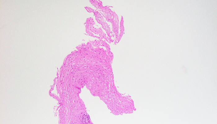
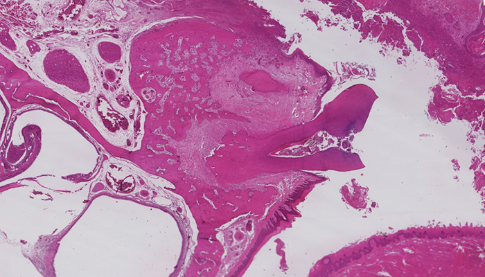
Keratin pearl in a tongue squamous cell carcinoma.
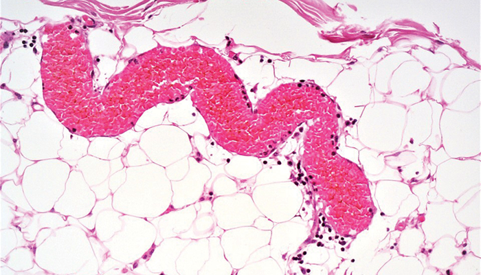
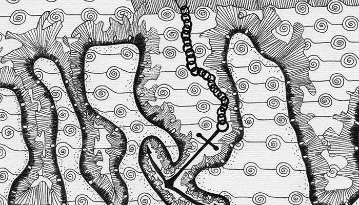
Adipose tissue and a snake-shaped blood vessel.
Amber Matkowski
Credit: Amber Matkowski, Junior Doctor, Wye Valley NHS Trust, UK.
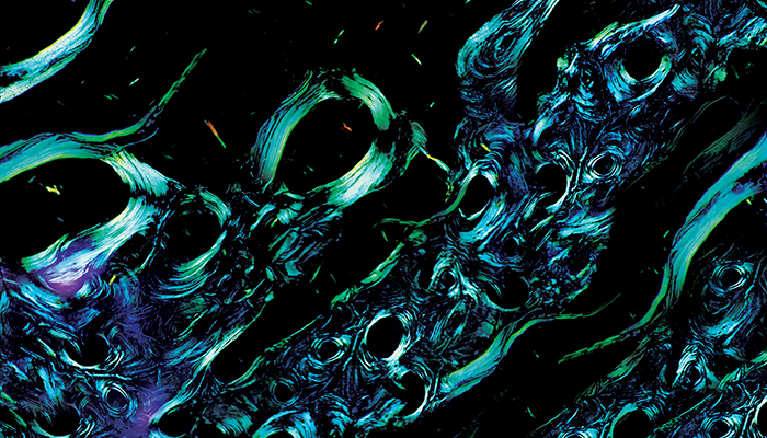
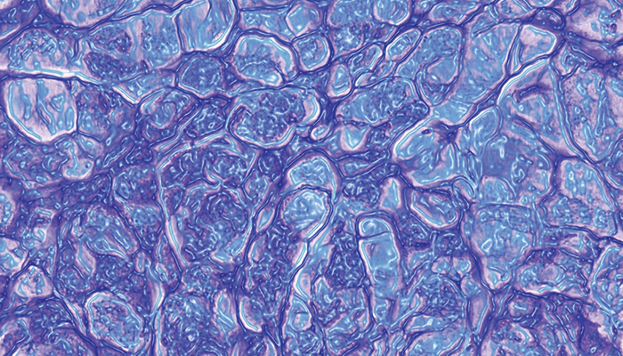
Digital manipulation of amyloid deposits visualized by polarized light microscopy.
Ana I. Hernandez Caballero
Credit: Ana I. Hernandez Caballero, Department of Pathology and Laboratory Medicine, University of Rochester Medical Center, Rochester, New York, USA
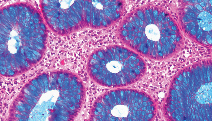
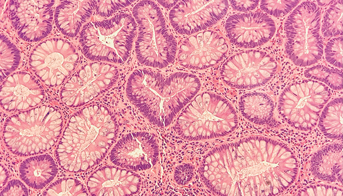
Kreyberg stain of a colon biopsy.
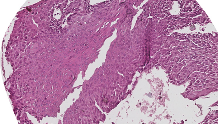
H&E stain of an esophageal biopsy.
B. Alan Rampy
Credit: B. Alan Rampy, Associate Professor of Diagnostic Medicine and Medical Education and Distinguished Teaching Professor, The University of Texas at Austin, Texas, USA.
A stylized example of histopathology from a mucinous cholangiocarcinoma.
Caroline Stanek
Credit: Caroline Stanek, PGY-1, Department of Pathology, Heersink School of Medicine, The University of Alabama at Birmingham, Alabama, USA
A tubular adenoma from a GI biopsy.
Christine Carreira
Credit: Christine Carreira, Research Assistant, WHO/IARC Classification of Tumours Group, International Agency for Research on Cancer, World Health Organization, Lyon, France.
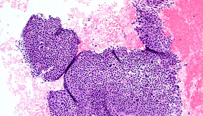
Human cervical biopsy CIN3.
Human cervical biopsy CIN3.
Cooper Schwartz
Credit: Cooper Schwartz, Warren Alpert Medical School of Brown University, Providence, Rhode Island, USA.
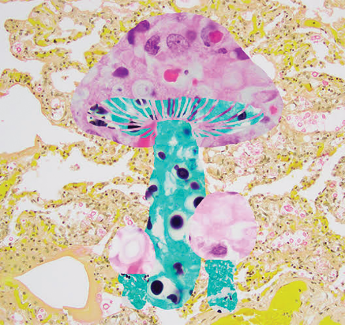
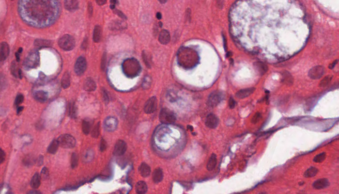
A collage of slides of lung tissue from a patient with disseminated Cryptococcus neoformans fungal infection.
Dalila Villalobos
Credit: Dalila Villalobos, PGY-5 Anatomical Pathology, Queen's University, Kingston, Ontario, Canada
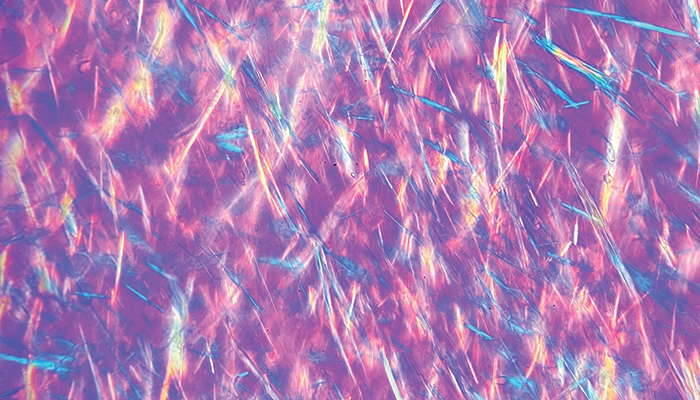
A needle-shape negative birefringent image of gout.
Emanuela Veras
Credit: Emanuela Veras, Highlands Pathology, Bristol, Tennessee, USA.
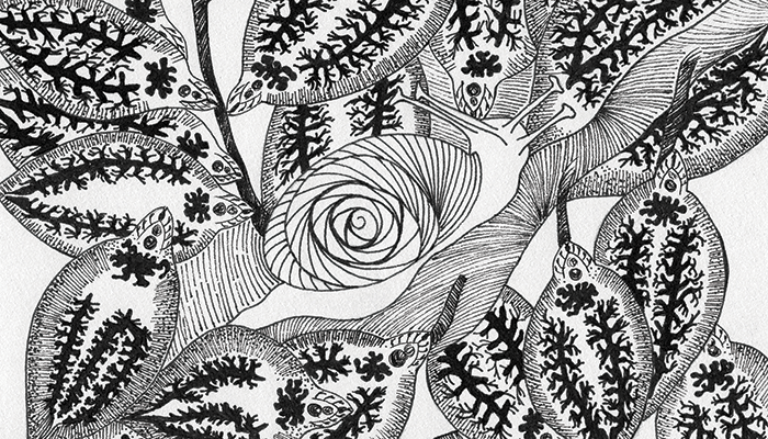
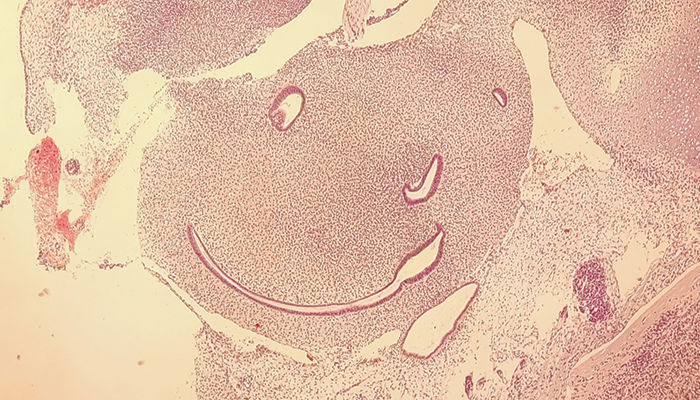
Pathology images are often reminiscent of real-life objects, animals, and many other things we frequently find in our daily surroundings. I am inspired by what I see under the microscope to create a connection between these two worlds.
Franz Jobert L. Sebastian
Credit: Franz Jobert L. Sebastian, Philippine Heart Center, Quezon City, Philippines.
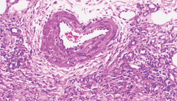
A long time ago in a galaxy far, far away... a galactic strike ship from an unknown planet is ready to invade and conquer the universe. This photomicrograph is a mitral valve from a patient with rheumatic heart disease with amyloid accumulation, stained with Congo red to show the characteristic apple-green birefringence on a polarizing microscope.
Frazer Bell
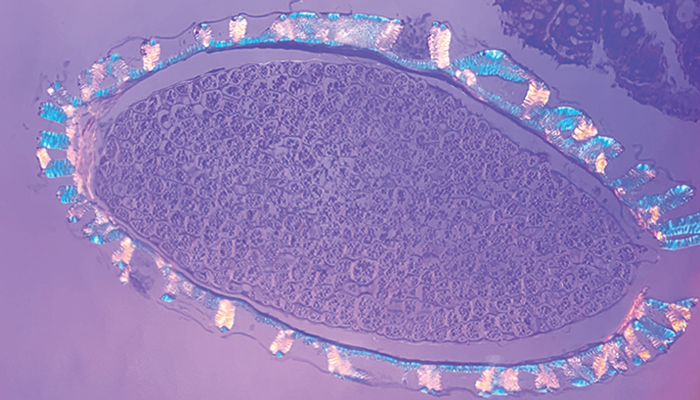
Credit: Frazer Bell, Histopathology Technician, Veterinary Diagnostic Services, University of Glasgow, Scotland, UK
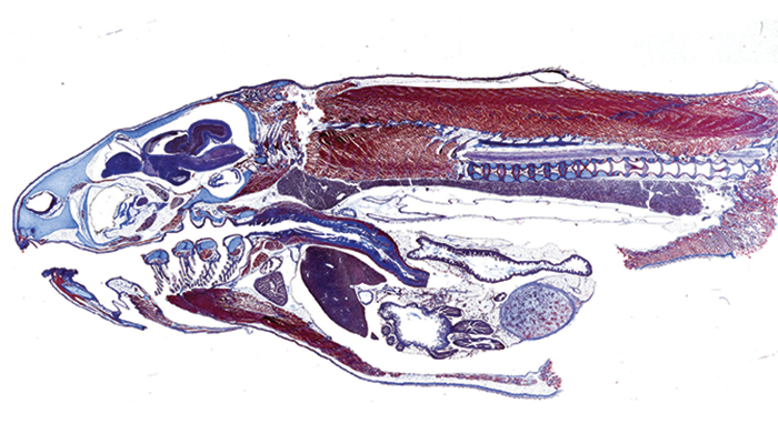
Longitudinal cross section of fish fry stained using Masson’s trichrome protocol. By Frazer Bell, Lynn Stevenson, Lynn Oxford, and Jan Duncan.
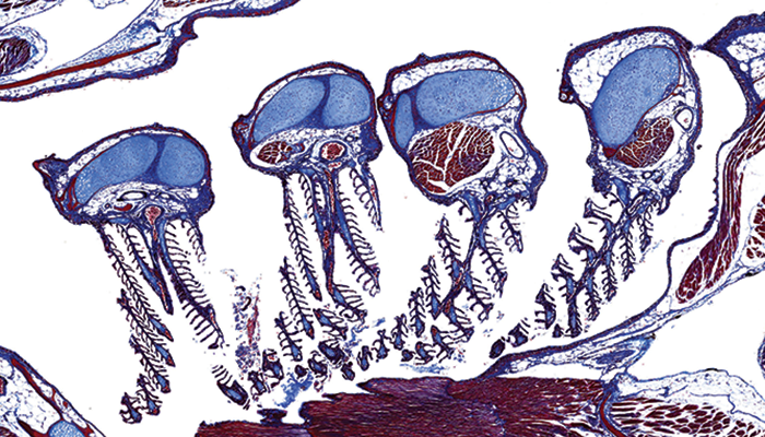
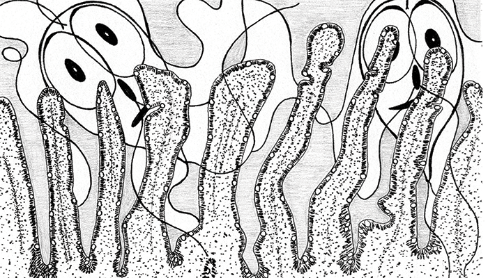
Fish gills looking like jellyfish during the medusa phase, stained using Masson’s trichrome protocol. By Frazer Bell, Lynn Stevenson, Lynn Oxford, and Jan Duncan.
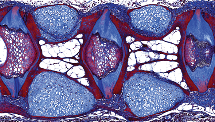
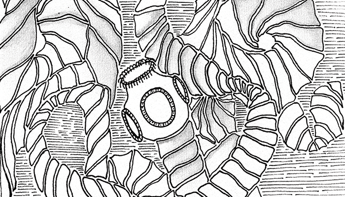
A magnified look into the spine of a fish, looking almost like a stained glass window. By Frazer Bell, Lynn Stevenson, Lynn Oxford, and Jan Duncan.
Angie Rupp
Credit: Angie Rupp, Senior Veterinary Clinician, Veterinary Diagnostic Services, University of Glasgow, Scotland, UK
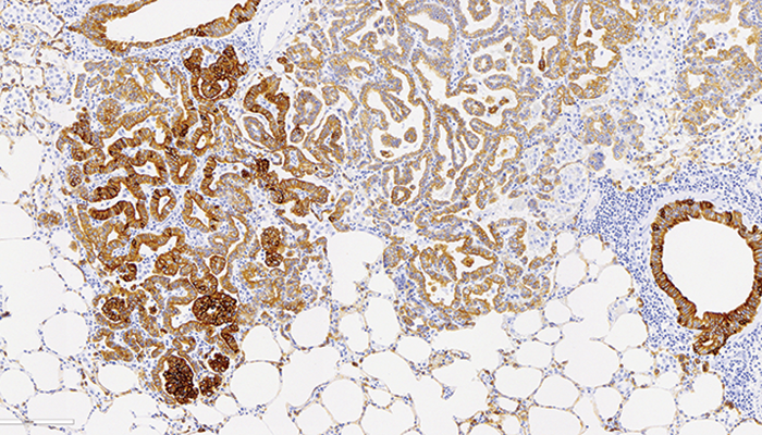
Ovine lung stained using IHC pan-cytokeratin. Ovine pulmonary adenocarcinoma (OPA) caused by jaagsiekte sheep rotavirus (JSRV). By Angie Rupp, Frazer Bell, Lynn Stevenson, Lynn Oxford, and Jan Duncan.
Gabriel Arismendi-Morillo
Credit: Gabriel Arismendi-Morillo, Pathologist, Professor, and Researcher, Electron Microscopy Laboratory, Biological Researches Institute, Faculty of Medicine, University of Zulia, Maracaibo, Venezuela.
Picture from a surgical piece with breast ductal carcinoma. (Histology technicians: G. Arteaga and P. Arteaga.)
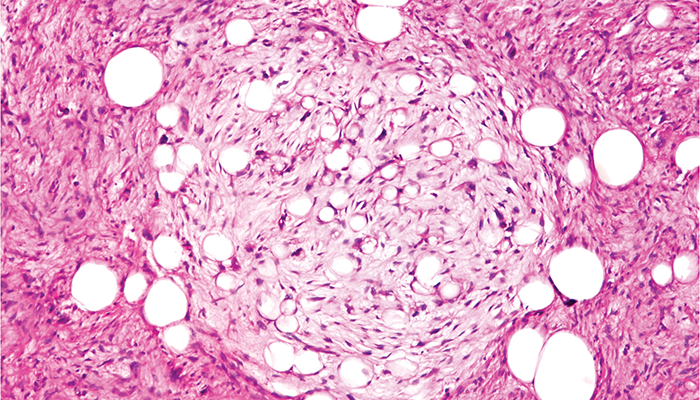
Picture from a chest tumor: pleomorphic liposarcoma. (Histology technicians: G. Arteaga and P. Arteaga.)
Picture from a brain tumor: pleomorphic xanthoastrocytoma. (Histology technicians: G. Arteaga and P. Arteaga.)
Mercia Locke
Credit: Mercia Locke, Cytotechnologist, Southern Arizona VA Health Care System, Tucson, Arizona, USA
When you're trying to screen slides, but pareidolia takes over...
Georgios Kitsakis
Credit: Georgios Kitsakis, Histopathology Resident, General Hospital of Volos, Greece.
Fetal tissue.
Hansini Laharwani
Credit: Hansini Laharwani, PGY-4 Pathology Resident, University of Mississippi Medical Center, Jackson, Mississippi, USA.
A polarized image of feces under a microscope.
Inny Busmanis
Credit: Inny Busmanis, Senior Consultant Pathologist, Singapore General Hospital, Singapore.
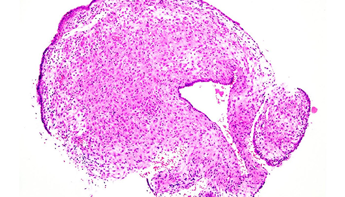
An incidental finding from an affectionate specimen. Taken using the InfinityCapture system.
Jagmohan S. Sidhu
Credit: Jagmohan S. Sidhu, Medical Director and Chairman, Department of Pathology and Laboratory Medicine, United Health Services Hospitals-Wilson Medical Center/ Binghamton General Hospital, Johnson City/Binghamton, New York, USA.
James Lewis, Jr.
Credit: James Lewis, Jr., Professor and Chief of Head and Neck and Endocrine Pathology, Vanderbilt University Medical Center, Nashville, Tennessee, USA
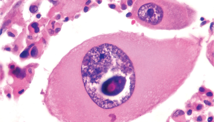
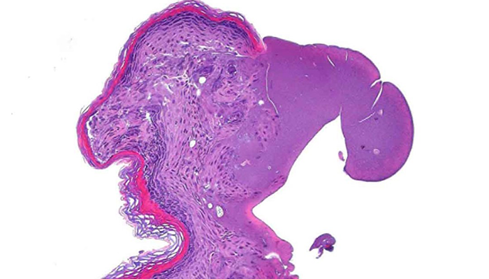
A most sarcastic oncocytic papilloma!
Jason Innerhofer
Credit: Jason Innerhofer, Lead Pathologists’ Assistant; Faculty Preceptor, Rosalind Franklin University; and Clinical Instructor, University of New Mexico, Albuquerque, New Mexico, USA.
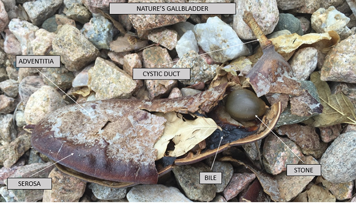
Pathology is in the eye of the beholder…
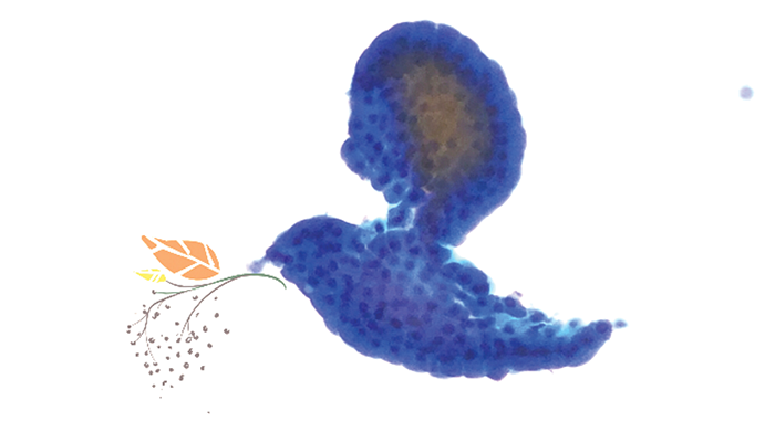
Jayashree Kulkarni
Credit: Jayashree Kulkarni, Consultant and Head of Hematopathology, Sri Shankara Cancer Hospital and Research Center, Bangalore, India.
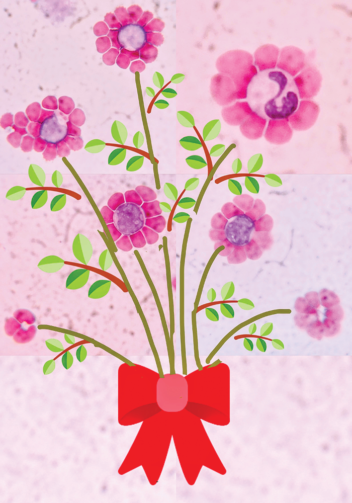
Cerebrospinal fluid light microscopy showing erythrocytes rosetting around lymphocytes and neutrophils. The bouquet was made using a grid art app.
José Antonio Ortiz Rey
Credit: José Antonio Ortiz Rey, Hospital Álvaro Cunqueiro, Vigo, Spain.
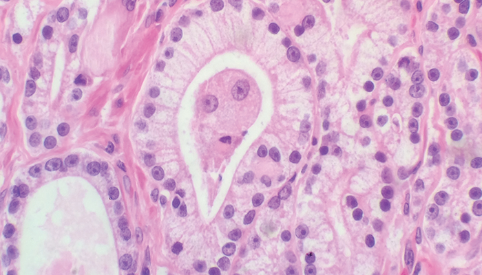
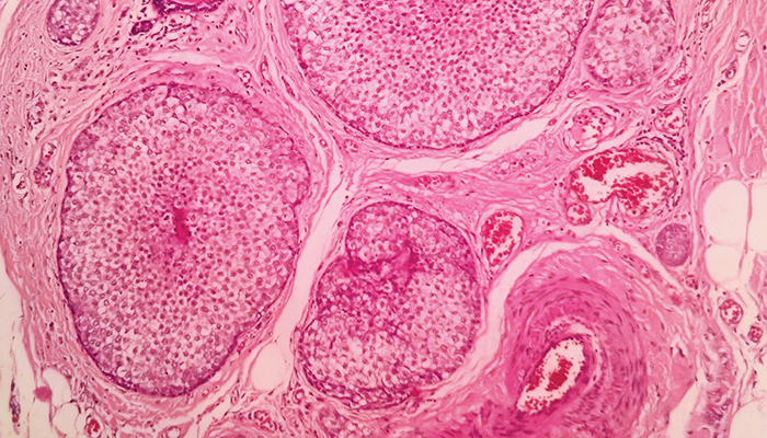
The Astonished Prostate Adenocarcinoma
Maaia Jentus
Credit: Maaia Jentus, Medical University of Vienna, Austria
People who have met me know that medicine is my biggest passion – a fire that burned even brighter when I met my pathology squad. It’s about the beauty of nature and mechanisms. About asking not “why?” but “how?” About endless hours spent sitting at a microscope, with whole-slide images on a screen, in the lab, in dissection rooms, listening to lectures and congresses… And now, I also help to improve pathology teaching. Curiosity makes science fun.
In 2017, I began thinking about future imaging possibilities. These colleagues are sitting around a table with a 3D holographic biopsy/MRI molecular image…
This art was a recent commission for a kidney pathologists as a present from his cardiologist wife.
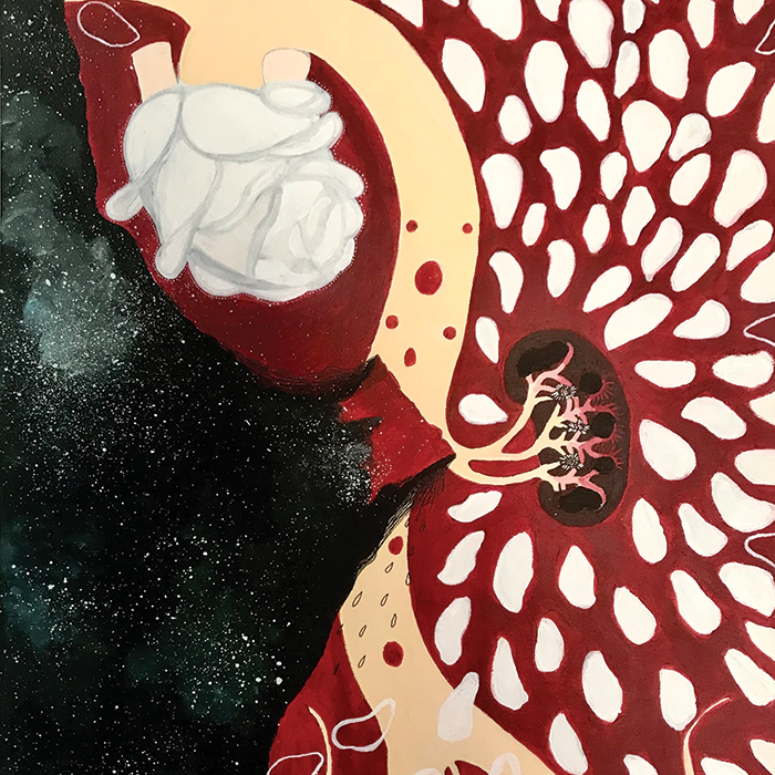
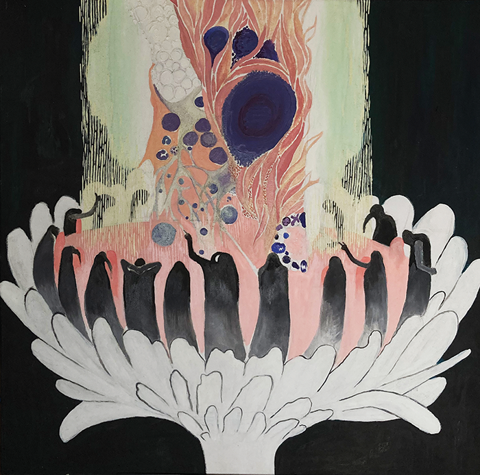
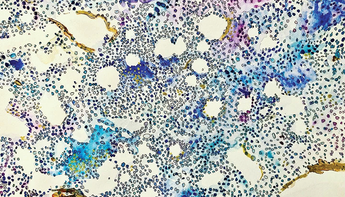
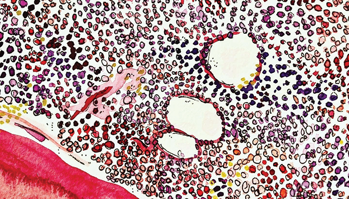
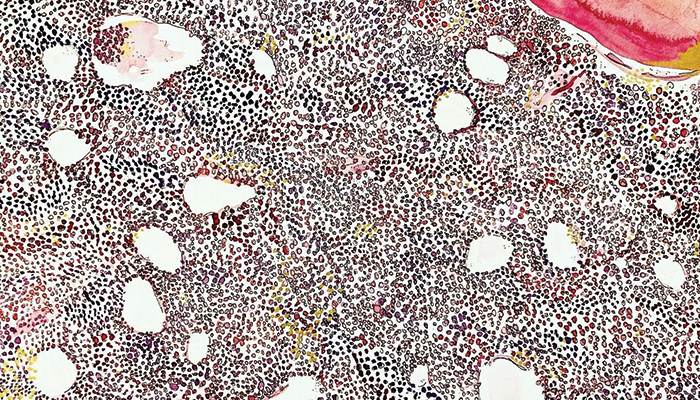
Yeonsoo Sara Lee
Credit: Yeonsoo Sara Lee, Resident, Department of Pathology, LAC+USC Medical Center, Los Angeles, California, USA
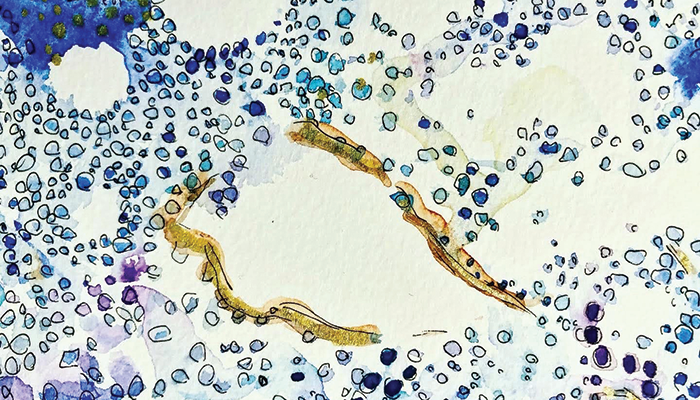
These pieces were created for a mentor of mine who is a pathologist. They are inspired by CD34 and H&E stains of bone marrow; the medium is watercolor, metallic paints, and ink. Though I am still open to the specialty I will be pursuing, I have a particular fondness for pathology and laboratory-based medicine. I was a field ecologist in a previous life (as a nontraditional medical student) and spent much time peering through scopes at different freshwater microorganisms. Disease ecology is eventually what brought me to medicine, so I am grateful for my time in the lab.
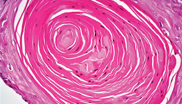
Mayah Hijazi
Credit: Mayah Hijazi, Resident, Department of Pathology, LAC+USC Medical Center, Los Angeles, California, USA
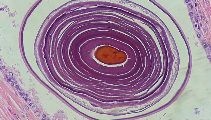
Gorgeous concentric circles in a corpus amylaceous. In reality, this would be a sphere; let your imagination run wild to this artistry in life.
Michael J. Klein
Credit: Michael J. Klein, Professor of Pathology and Laboratory Medicine at Weill-Cornell College of Medicine and Pathologist in Chief Emeritus at the Hospital for Special Surgery, New York, USA
At the Hospital for Special Surgery, we were lucky enough to be able to use whole-slide imager (with FDA approval) to do at least some of our signout work from home.


Nika Gloyeske
Credit: Nika Gloyeske, Clinical Assistant Professor, Pathology and Laboratory Medicine, The University of Kansas Health System, Kansas City, Kansas, USA
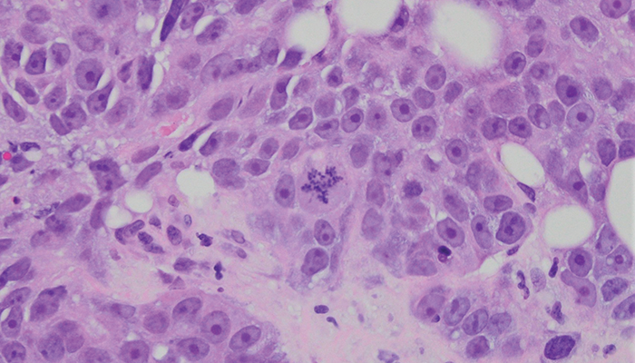
A uniquely shaped mitosis in a triple-negative breast cancer.
Nusaiba Alrefae
Credit: Nusaiba Alrefae, Cytopathology Unit, Sabah Hospital, Kuwait
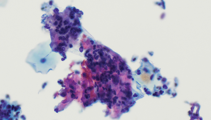
An image taken from ThinPrep preparation of a fine needle aspiration biopsy of a thyroid nodule.
Richard Prayson
Credit: Richard Prayson, Cleveland Clinic, Cleveland, Ohio, USA.
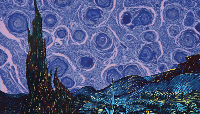
Psammomatous meningioma in the background sky of Vincent van Gogh’s Starry Night.
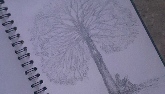
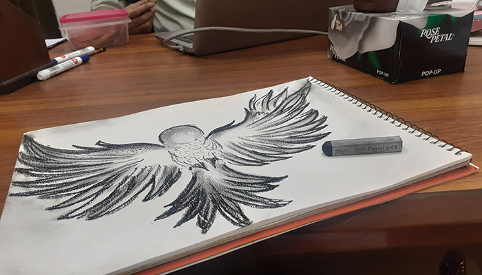
Sadaf Sarfraz
Credit: Sadaf Sarfraz, Molecular Pathology and Genomics, Forman Christian College University, Lahore, Pakistan.
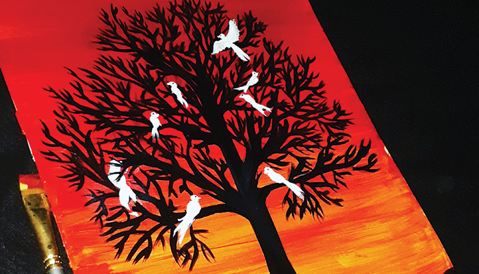
A collection of paintings by cancer researcher Sadaf Sarfraz.
Sanjay A. Pai
Credit: Sanjay A. Pai, Consultant Pathologist and Head of Pathology, Manipal Hospital Yeshwanthpur, Bangalore, India.
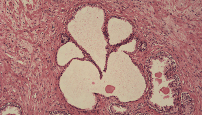
This image of a prostatic gland has an uncanny resemblance to Lord Ganesha in Hindu religion and mythology. Note that Lord Ganesh often has a rat near his feet – and even that is depicted in this prostate!
Sheryl Lammers
Credit: Sheryl Lammers, Retired Medical Technologist, Metropolitan Medical Laboratory, Moline, Illinois, USA
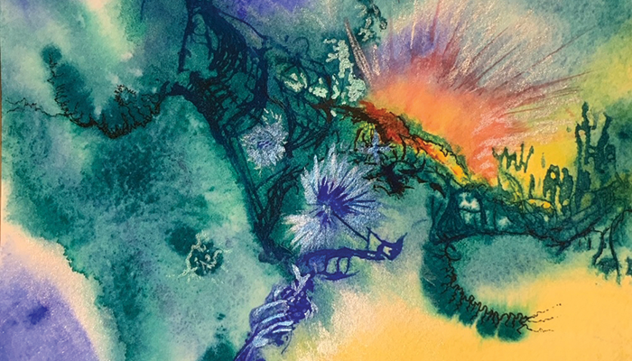
Watercolor and ink.
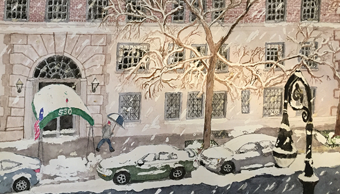
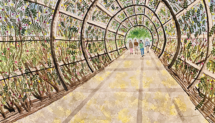
Supriya R. Donthamsetty
Credit: Supriya R. Donthamsetty, Ochsner Medical Center, New Orleans, Louisiana, USA.
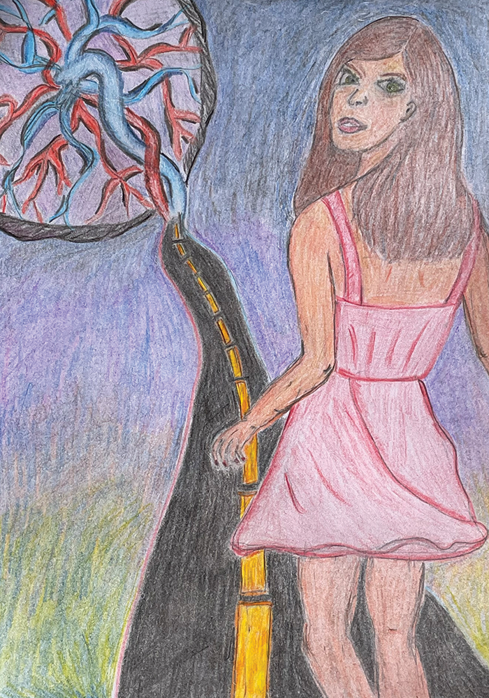
A young woman is walking along a road and looking back with uncertainty. The end of the road turns into an umbilical cord and ends in a placenta. The placenta symbolizes motherhood; the road the connection of the young woman with her mother. It is uncertain why this young woman is seeking connection with her mother at this particular time in her life.
Newsletters
Receive the latest pathologist news, personalities, education, and career development – weekly to your inbox.


