
Esophageal adenocarcinoma (EAC) is one of the deadliest cancers in the US, and Barrett’s esophagus (BE) is its sole precursor. Most patients with BE have non-dysplastic BE (NDBE) and are at low risk of developing EAC. However, some patients with NDBE are at high risk of progression, and traditional management is largely unable to identify these high-risk individuals.
Robert Najarian, an anatomic pathologist and leading expert in gastrointestinal pathology, sat down with us to discuss this challenge and why he advocates for use of TissueCypher to risk-stratify patients with NDBE.

From a pathologist’s perspective, can you describe the challenge of identifying which patients with NDBE are likely to develop EAC?
BE is relatively common in the US, with about one in 15 individuals having the condition. Approximately 90 percent of individuals with BE have NDBE, which is characterized by presence of intestinal metaplasia but lacks overt signs of dysplasia.
As most patients with NDBE will not progress to EAC, gastroenterology society guidelines recommend that they undergo surveillance endoscopy every 3 to 5 years and have biopsies from those endoscopies reviewed by pathologists such as myself to detect any progression toward EAC.
Despite the low overall risk of progression of NDBE, a subset of patients with NDBE are at high risk of progression and may develop EAC before their next guideline-recommended endoscopy. There is little that we, as pathologists, can do to figure out who the high-risk NDBE patients are.
Why is the traditional approach unable to identify patients with NDBE at high risk of developing EAC?
The traditional approach to risk-stratifying patients with BE primarily relies on the patient’s histologic grade, and their BE segment length if they have NDBE. For patients with NDBE, these factors aren’t sufficient, as 50 percent of patients that progress to high-grade dysplasia (HGD) or EAC have an index pathological assessment of NDBE.
Those patients with NDBE at high risk of progressing to HGD or EAC may lack clear signs of dysplasia. As a result, if I find that a patient has NDBE, there is no way for me or the managing physician to clearly know whether the patient is truly at low risk of progression to EAC. This is the crux of the problem.
What can pathologists do to help clinicians risk-stratify patients with NDBE?
NDBE lacks visible signs of dysplasia, but a patient’s risk of neoplastic progression can be assessed by means other than histologic review. Molecular tests are designed to detect the biological changes that precede signs of dysplasia and ultimately drive progression to EAC. Pathologists can use and advocate for these tests to augment our ability to equip clinicians with prognostic information that histology cannot provide.
A common adjunctive tool is p53 immunohistochemistry (IHC), which is accessible to pathologists and may provide added diagnostic confidence to help remove ambiguity surrounding a histological assessment of low-grade dysplasia (LGD). From a prognostic perspective, though, p53’s use as a risk stratification tool is highly variable and subjective.
Other approaches, including assessment of copy number variations and use of fluorescence in situ hybridization (FISH), are also available to pathologists, but they lack extensive evidence as to their clinical validity and utility and require specialized equipment to perform the staining and manual, visual interpretation. TissueCypher (TSP-9) is the most extensively validated and commercially available approach to risk-stratify patients with NDBE.
What is TissueCypher and what led you to advocate for its use to risk stratify patients with NDBE?
TissueCypher is a tissue systems pathology test that objectively risk stratifies patients with BE, including NDBE, indefinite for dysplasia (IND), and LGD, for progression to HGD/EAC. The test uses sections from routine pinch biopsy specimens to perform multiplexed immunofluorescent labeling of nine protein biomarkers, including p53, in seven cell and tissue structures to quantify 15 spatialomic features within each BE specimen. These features are then integrated into a locked, AI-derived risk classifier to determine a patient’s risk score, a risk class, and 5-year probability of progression to HGD/EAC (Figure 1).
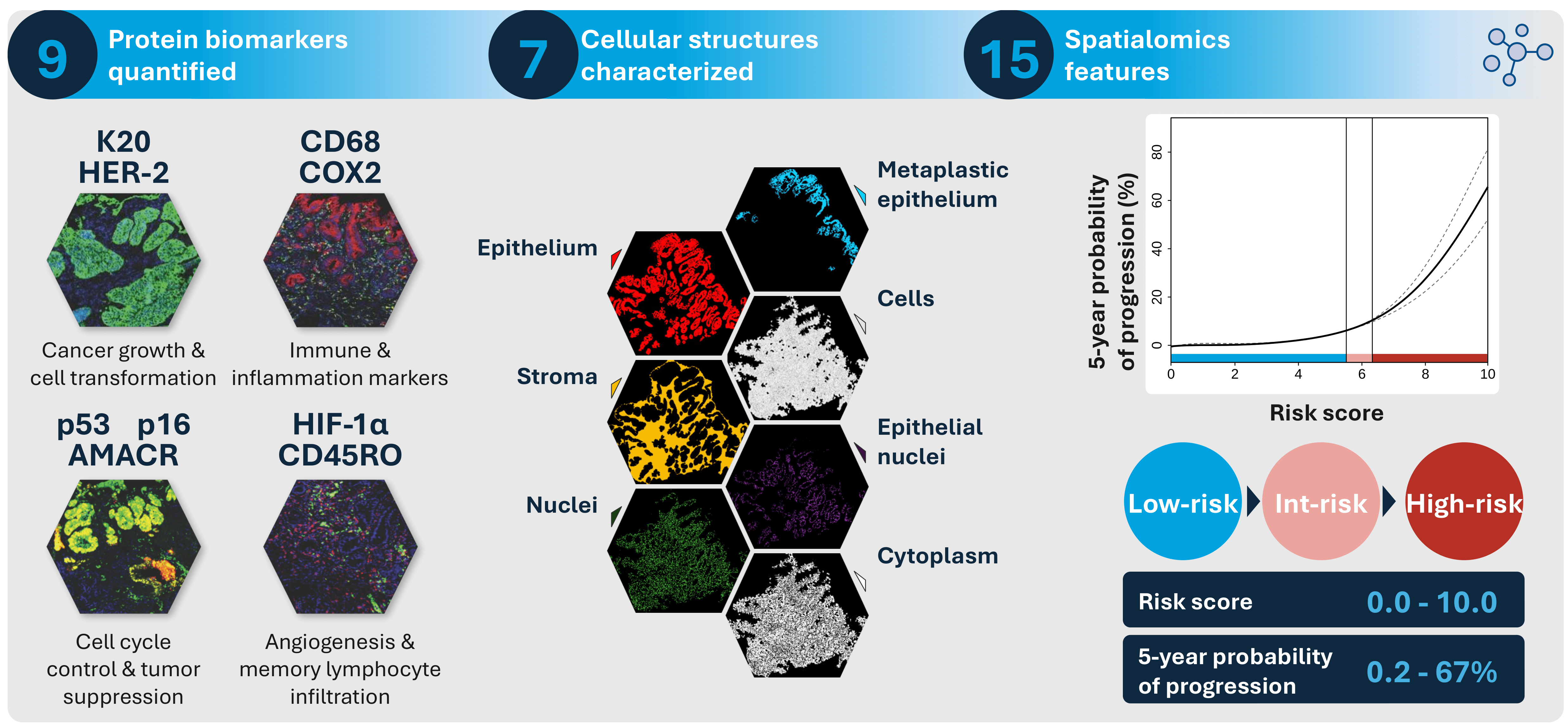
The ability of TissueCypher to help identify which patients with NDBE are at high or low risk of progression to HGD or EAC is what led me to advocate for use of the test. The test has also been validated across multiple clinical studies, has demonstrated improved sensitivity and specificity over pathology-only risk assessment, and has been shown to be the strongest independent predictor of progression.
TissueCypher is also designed to reduce the variability associated with sample staining and interpretation that can confound risk stratification by other approaches such as p53 IHC. Moreover, TissueCypher is trusted by many of my pathologist and GI physician colleagues.
Are there any closing thoughts you’d like to share?
Though I am not in a patient-facing role, the nature of my work affords me the privilege to directly impact care for many patients across the country. Patients with NDBE have not traditionally had many options for precision risk assessment and early intervention until TissueCypher became clinically available. Assessing multiple biomarkers is a powerful approach to informing personalized, risk-aligned management of patients with NDBE whose risk of progression can be unclear via histology.
Pathologists should feel empowered to work closely with gastroenterologists and advanced practice providers to discuss tools like TissueCypher to improve care for patients with BE. Pathologists don’t always use the power they have in their role as experts in diagnostic medicine, but, armed with strong data and a passion to enact change, they can be a critical advocate and resource to improve patient care.
Newsletters
Receive the latest pathologist news, personalities, education, and career development – weekly to your inbox.