Markku Martti Miettinen is a Finnish-born surgical pathologist and senior clinician at the National Cancer Institute (NCI), National Institutes of Health (NIH), where he heads the General Surgical Pathology Section in the Laboratory of Pathology. He is widely considered to be the leading authority on gastrointestinal stromal and soft tissue tumors and serves as a consultant to surgical pathologists in the United States and worldwide.
He has authored more than 500 peer-reviewed articles, which have been cited over 73,000 times, with a h-index of 136. These metrics place him among the most cited pathologists globally.
Here, Miettinen joins Ivan Damjanov to discuss his exemplary career in the lab and beyond.
Where did your interest in medicine and pathology come from?
My initial plan was to become a botanist. During my school years, I was fascinated by flora and fauna and regularly took part in activities with my school’s Nature Club, which reinforced my interest in botany.
However, a few months before graduating from high school, I changed course and applied to medical school in Helsinki. My decision was h4ly supported by my grandmother, who had previously hoped my father would become a doctor. When he chose to study law instead, she turned her encouragement toward me.
I later decided to pursue pathology after being inspired by my professor, Erkki Saxén. I still remember how captivated I was by his lectures and by viewing tumor slides, even though the projections – produced with an old microscope using carbon electrodes – were far from impressive by today’s standards.
Further inspiration came from spending two summer vacations at a small country hospital, where a practicing pathologist taught me how to perform autopsies and introduced me to diagnostic surgical pathology under the microscope. I actually began my formal training there, post graduation, after finding that all residency positions at the University of Helsinki were filled.
However, six months later, I received an unexpected call from the university offering a temporary residency position while another resident was on maternity leave. I accepted and joined the program, which set me firmly on the path to becoming a pathologist. Returning to Helsinki also allowed me to reunite with my girlfriend, whom I married soon afterward.
How did you decide to move to the US?
Immediately after graduating from medical school, I took the American licensing examination administered by the Educational Council for Foreign Medical Graduates (ECFMG). I do not clearly remember why, as I had no plans to move to the US at the time. The exam, held at the US Embassy in Helsinki, consisted of a medical component and a spoken English test. I passed the medical portion but failed the English section because I did not understand all the words delivered through headphones. At school, I had focused on Latin, so my English was limited.
As I began my pathology training, however, I realized that most scientific literature was published in English. I therefore made a serious effort to improve my language skills and later passed the English component of the ECFMG exam.
While completing my doctoral thesis on lymph node toxoplasmosis and related lymphadenopathies, I attended an international conference in Hamburg, Germany, where we met for the first time.
You encouraged me to apply for a postdoctoral position at Hahnemann University in Philadelphia. I accepted and moved there with my wife and children. We stayed for one year. Although I was offered the opportunity to remain, I was not ready to settle in the US, and we returned to Finland, where I continued my clinical work and research in Helsinki.
My hospital workload in Helsinki was heavy, and I often conducted research in the evenings. After dinner with my family, I would frequently return to the pathology department and work late into the night, sometimes taking the last city bus home at 2am.
One evening, you called me at the laboratory. You were moving with your chairman, Emanuel Rubin, to Thomas Jefferson University in Philadelphia and invited me to join your team. Having previously worked with you, I accepted the offer without hesitation. With my wife’s support, we moved with our three children to Philadelphia.
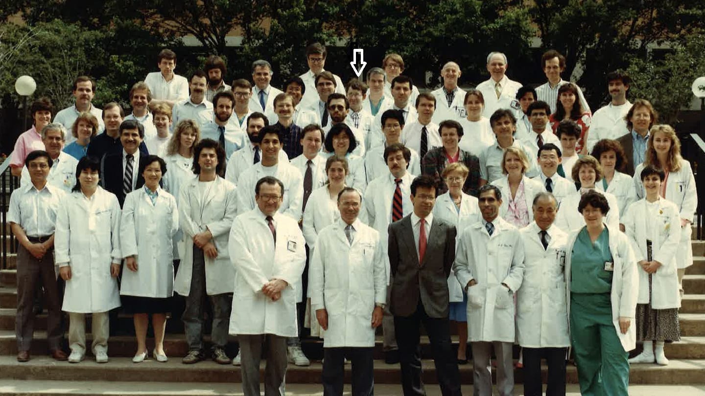
There, I joined a collegial and supportive department of 54 staff members. Under Rubin’s leadership, the department was ranked fifth among NIH-funded institutions, providing an excellent environment in which to begin my career in the US.
As a board-certified pathologist, you worked at Jefferson Medical College and later at the Armed Forces Institute of Pathology (AFIP). What were the key milestones of your career at that time?
At Jefferson Hospital, I learned automated immunohistochemistry and diagnostic hematopathology, which I practiced actively at the time. I also began collaborating with an experienced soft tissue and bone surgeon. During this period, I met Jerzy Lasota, a Polish scientist with advanced expertise in molecular biology. When I moved to the AFIP in 1996, I encouraged them to join me. We later moved together to the National Institutes of Health (NIH) and have continued our collaboration ever since.
At AFIP, I had more time to focus on research because I was no longer burdened with routine hospital duties. This was an exciting period in my career. I benefited from access to world-class pathology archives, h4 support from colleagues, and a highly collaborative environment. As a result, my research productivity increased, and I established several international collaborations.
During this time, I developed a h4 interest in gastrointestinal stromal tumors (GIST). Together with Lasota, I began collaborating with Leslie Sobin, Chief of the Gastrointestinal Branch at AFIP. This work became a major focus of my research and led to my involvement in the international “GIST study group,” which was gaining significant attention at the time. Although GISTs are relatively rare, the scientific interest in them – particularly as a model for targeted therapies – allowed us to assemble a large cohort of cases, including 1,765 gastric and 906 small intestinal tumors, as well as cases from other sites.
In addition, access to the AFIP archives enabled me and my collaborators to define several new soft tissue tumor entities, including sclerosing perineurioma, lipofibromatosis, gastroblastoma, and plexiform fibromyxoma of the stomach.
What are your current responsibilities at the NIH?
I am a senior clinician and principal investigator, which means I lead a funded research program that is reviewed every four years by an external panel at the NIH to ensure accountability.
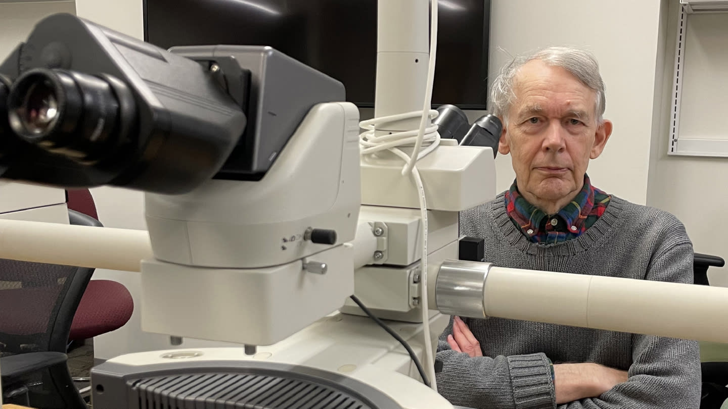
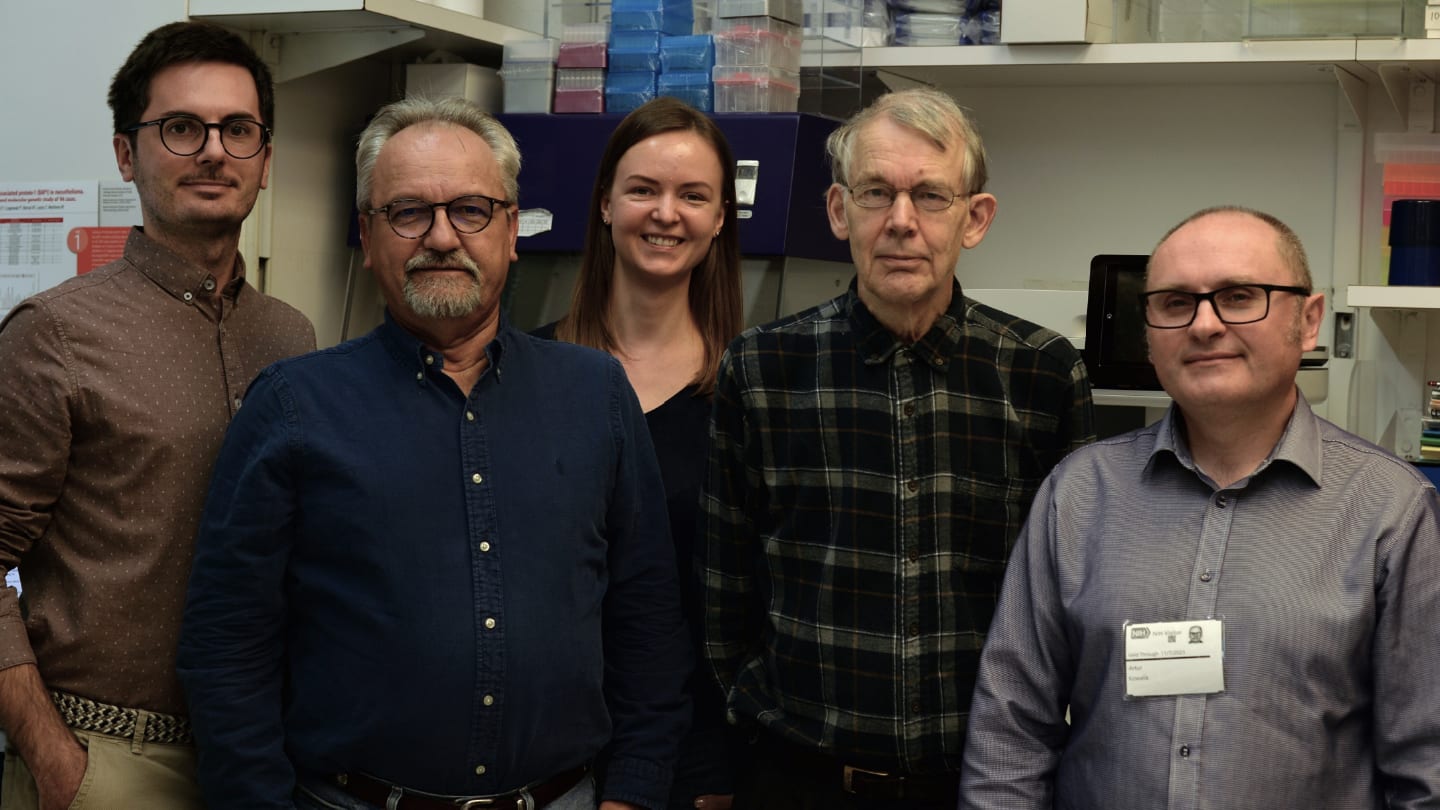
I oversee surgical pathology within a team of five pathologists, each with different areas of expertise. I also maintain an active consultation practice, with our laboratory receiving slides from several thousand cases for second opinions.
In addition, I give seminars and presentations in the US and internationally when my clinical and research responsibilities allow.
Can you tell us about some of your most cited articles?
Certainly! The paper “Gastrointestinal stromal tumors: definition, clinical, histological, immunohistochemical, and molecular genetic features and differential diagnosis” (Virchows Archiv, 2001), cited 3,250 times, is a seminal review that helped define GIST as a distinct tumor entity following the discovery of KIT mutations. It provided a clear framework for distinguishing GISTs from true smooth muscle tumors and has been highly influential in shaping modern diagnostic practice.
Another key publication, “Gastrointestinal stromal tumors: pathology and prognosis at different sites” (Seminars in Diagnostic Pathology, 2006), cited 2,615 times, established the concept that GIST behavior varies by anatomical site. This work highlighted important differences between tumors arising in the stomach, small intestine, and rectum, and remains central to current approaches to risk assessment and staging.
And finally, “KIT (CD117): a review on expression in normal and neoplastic tissues, and mutations and their clinicopathologic correlation” (Applied Immunohistochemistry & Molecular Morphology, 2005), cited 1,170 times, is an important reference on the diagnostic use of CD117. It explains its role in identifying GIST as well as its expression in other tumor types, providing essential context for accurate interpretation in pathology practice.
What are some of the scientific problems you’re currently tackling?
My current projects cover diagnostic improvement of lipomatous tumors and fusion sarcomas. My laboratory also studies diagnostic and actionable tumor markers using immunohistochemistry to develop new tools for our oncology colleagues in patient care.
Newsletters
Receive the latest pathologist news, personalities, education, and career development – weekly to your inbox.


