Conexiant
Login
  • The Analytical Scientist
  • The Cannabis Scientist
  • The Medicine Maker
  • The Ophthalmologist
  • The Pathologist
  • The Traditional Scientist
The Pathologist
  • Explore Pathology

    Explore

    • Latest
    • Insights
    • Case Studies
    • Opinion & Personal Narratives
    • Research & Innovations
    • Product Profiles

    Featured Topics

    • Molecular Pathology
    • Infectious Disease
    • Digital Pathology

    Issues

    • Latest Issue
    • Archive
  • Subspecialties
    • Oncology
    • Histology
    • Cytology
    • Hematology
    • Endocrinology
    • Neurology
    • Microbiology & Immunology
    • Forensics
    • Pathologists' Assistants
  • Training & Education

    Career Development

    • Professional Development
    • Career Pathways
    • Workforce Trends

    Educational Resources

    • Guidelines & Recommendations
    • App Notes
    • eBooks

    Events

    • Webinars
    • Live Events
  • Events
    • Live Events
    • Webinars
  • Profiles & Community

    People & Profiles

    • Power List
    • Voices in the Community
    • Authors & Contributors
  • Multimedia
    • Video
    • Pathology Captures
Subscribe
Subscribe

False

The Pathologist / Issues / 2025 / October / Mapping the Brain’s Hidden Nanotube Network
Neurology Microscopy and imaging Digital Pathology

Mapping the Brain’s Hidden Nanotube Network

New imaging shows neurons may share signals and disease-related molecules through a previously unseen system of nanotubes

10/30/2025 News 1 min read

Share

Credit: Adobe Stock

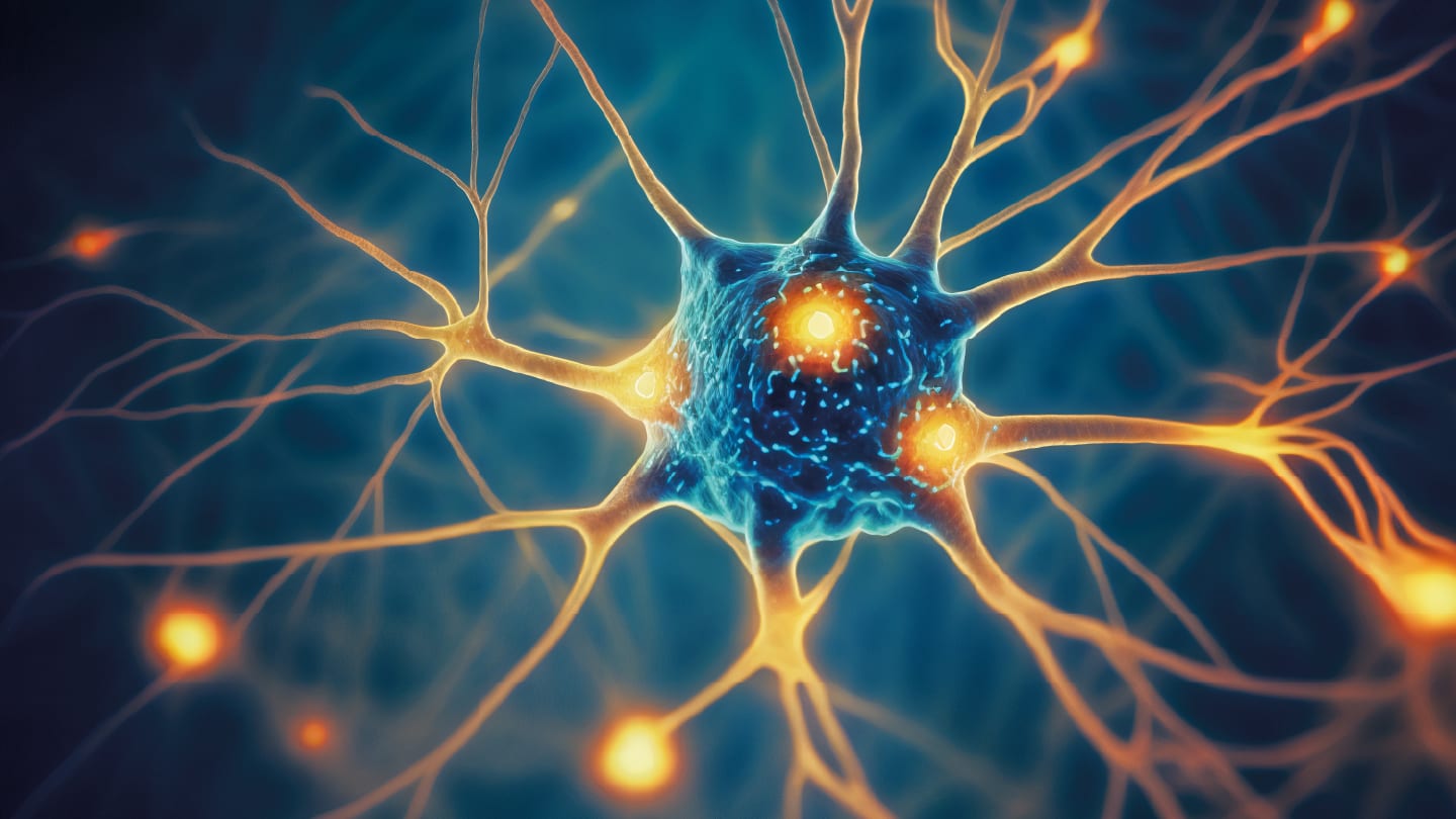
A study published in Science describes a new way neurons may communicate – through dendritic nanotubes (DNTs), ultrathin bridges that link cells outside traditional synapses. The discovery adds a potential new layer of connectivity in the brain and may help explain how disease-related molecules spread between neurons.

Neuronal communication has long been attributed to synapses, the specialized junctions that pass electrical and chemical signals. However, researchers observed membrane-to-membrane contacts between neighboring dendrites that lacked the typical features of synapses. Using super-resolution microscopy and computational image analysis, they confirmed these structures as distinct.

The DNTs form physical channels that allow the exchange of calcium ions and other molecules directly between neurons. When their formation was experimentally blocked, this nonsynaptic transfer stopped – demonstrating that DNTs provide a functional route for intercellular signaling.

The team tested whether these nanotubes could move disease-associated materials. When β-amyloid (Aβ) peptides, a hallmark of Alzheimer’s disease (AD), were injected into individual neurons in mouse brain tissue, the peptides traveled to neighboring cells through DNTs. Inhibiting nanotube formation halted the spread, suggesting that DNTs can act as conduits for pathogenic proteins.

In a mouse model of AD, DNT networks showed structural changes early in disease development – before visible amyloid plaque formation. Computational simulations supported these findings, predicting that overactive nanotube signaling could contribute to localized Aβ buildup and neuronal stress. This points to a potential mechanism for how neurodegenerative pathology may propagate in its earliest stages.

The study highlights a previously unrecognized cellular structure that may influence disease progression and biomarker distribution in brain tissue. DNTs could become important in diagnostic research, helping to interpret how toxic proteins move and accumulate within neural networks.

By revealing this nonsynaptic communication system, the researchers expand the framework for studying how neurons share materials – and how disruptions in that process might signal or accelerate disease.

Newsletters

Receive the latest pathologist news, personalities, education, and career development – weekly to your inbox.

Newsletter Signup Image

Explore More in Pathology

Dive deeper into the world of pathology. Explore the latest articles, case studies, expert insights, and groundbreaking research.

False

Advertisement

Recommended

False

Related Content

Intense Intestines
Microscopy and imaging
Intense Intestines

January 19, 2024

1 min read

Some powerful fluorescent microscopy in this Image of the Month…

Context Matters in Cancer Biology
Microscopy and imaging
Context Matters in Cancer Biology

December 27, 2021

1 min read

Akoya is leading the way with spatial phenotypic signatures – a novel class of biomarkers for predicting response to immunotherapy

When Pathology Goes 3D
Microscopy and imaging
When Pathology Goes 3D

February 2, 2022

1 min read

Stratifying cancers with non-destructive 3D pathology

Sacrificing Safety for Speed?
Microscopy and imaging
Sacrificing Safety for Speed?

February 4, 2022

3 min read

Wide adaptation of antigen testing could increase the risk of exposure to infectious SARS-CoV-2 samples

False

The Pathologist
Subscribe

About

  • About Us
  • Work at Conexiant Europe
  • Terms and Conditions
  • Privacy Policy
  • Advertise With Us
  • Contact Us

Copyright © 2025 Texere Publishing Limited (trading as Conexiant), with registered number 08113419 whose registered office is at Booths No. 1, Booths Park, Chelford Road, Knutsford, England, WA16 8GS.