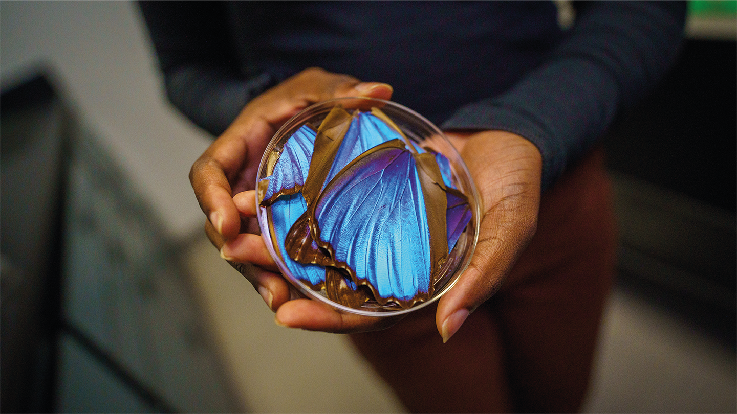
Scientists have been looking for the key to improved cancer diagnostics since its conception, but have we been searching in the wrong places? Researchers at the University of California San Diego have found success in taking inspiration from butterfly wings. Using the microscopic structures found on Morpho butterfly wings, the team have developed a simple and cost effective solution to analyze cancerous tissues. To learn more about this initiative, we spoke with Lisa Poulikakos, Paula Kirya, Jing Yang, and Aida Mestre-Farrera – the researchers behind this project.
What inspired this research?
Lisa Poulikakos was inspired by the important role that fibrous tissue microstructures play in the origin and progression of many serious diseases, including Alzheimer’s disease, heart disease, and various cancers. She learned that these tissues often have aligned protein structures, such as collagen, that interact with polarized light. However, detecting these interactions usually requires complicated and time-consuming staining methods or expensive imaging tools like Second Harmonic Generation microscopy, which are not widely accessible.
To solve this, the Poulikakos Lab began designing special photonic surfaces that enhance how polarized light interacts with tissues. This allows for clear, stain-free imaging of tissue structures using regular optical microscopes.
Paula Kirya, the lead author of this study, got involved after studying the shimmering colors of Morpho butterfly wings at Pasadena City College. These colors come from how light scatters off microscopic surface structures, not pigments. When she joined the Poulikakos lab, she noticed similarities between the lab's photonic surfaces and butterfly wings. This led her to suggest using actual Morpho wings to examine tissue microstructures.
Together, Kirya and Poulikakos discovered that the butterfly wings enhanced the visibility of unstained tissue samples. This breakthrough led to the creation of Morpho-Enhanced Polarized Light Microscopy (MorE-PoL), a new imaging platform.
Working with Jing Yang and Aida Mestre-Farrera from the UCSD School of Medicine, the team tested MorE-PoL on breast cancer biopsy samples with different collagen levels. Their results demonstrated the potential of this innovative, accessible imaging approach.
Did you face any challenges in the creation of this imaging technique, and if so, how did you overcome them?
One of the biggest challenges in developing the MorE-PoL imaging technique was figuring out how to accurately measure the images it produced. We wanted to turn the bright optical signals we saw into a mathematical model that could describe the amount and structure of fibers in tissue samples. To do this, we had to compare existing methods for analyzing optical materials with current ways of studying collagen fibers in tissue. The solution came through the interdisciplinary nature of our project – we combined knowledge from both optics and biology to find the right mathematical tools from optical physics to describe fibrotic tissue.
How does the MorE-PoL technique work?
The tiny scales on Morpho butterfly wings have fine ridges that act like a diffraction grating. This structure gives the wings a clear directional axis, making them optically anisotropic – in other words, how much light they reflect depends on the direction the light is coming from or the orientation of the wing.
This same optical anisotropy is also seen in tissue fibers like collagen, which are long, straight structures. These fibers also reflect polarized light differently depending on the direction – but their response is much weaker, making it hard to detect without special stains, equipment, or image processing.
To solve this, we place tissue samples directly on top of a section of Morpho butterfly wing. The wing boosts the anisotropic signal by nearly two times, making the tissue fibers easier to see under a microscope.
In making the imaging software, we cut out a piece of a preserved Morpho wing, attached it to a glass slide, and covered it with a coverslip. Then, we placed the tissue slide on top. This setup is imaged using a polarized light microscope with polarizers that are set at 90 degrees to each other – one polarizer allows only horizontal light in, and the other only allows vertical light out to the camera. When the tissue’s fibers are at a 45-degree angle, we get the strongest signal. When aligned with the polarizers, the image goes dark.
By rotating the microscope stage from 0 to 180 degrees, we measure how the brightness of the image changes. The difference between the brightest and darkest points tells us how many fibers are present (density), and how consistently the brightness changes tells us how well-aligned the fibers are (order). We use a mathematical tool called Jones Calculus to calculate both of these properties from the light patterns.

How does MorE-PoL compare to conventional histological techniques in identifying cancerous tissue?
H&E staining is the gold standard in pathology and helps pathologists reliably examine tissue. However, it doesn't provide enough information about the direction, amount, and arrangement of extracellular fibers – details that are important for diagnosing and predicting disease.
To get this extra information, pathologists often use special stains like Masson’s Trichrome or immunohistochemistry. But these techniques can introduce problems, such as inconsistent staining, personal interpretation bias, and long, tedious procedures.
With MorE-PoL, we take advantage of the natural physical properties of tissue to reveal its structure without using any stains. The process is stain-free, contact-free, and the Morpho butterfly wing section used for imaging can be reused over long periods. This makes it faster and more resource-efficient to analyze fiber organization in tissues, while avoiding the issues that come with traditional staining methods.
Could this technique be seamlessly integrated in standard pathology workflows?
This method can be used alongside H&E staining to give even more detailed information about the extracellular matrix, which is important for fully understanding disease. As our image analysis tools improve, we can speed up the process of identifying fiber patterns and linking them to disease outcomes.
In resource-limited settings, it’s often harder to study the structure of the extracellular matrix because it usually requires special stains or expensive imaging tools like multiphoton confocal microscopy, Second Harmonic Generation microscopy, or electron microscopy. MorE-PoL offers a low-cost, stain-free alternative that can provide similar insights. Plus, the Morpho butterfly wing can be reused many times to image multiple samples, making this a sustainable and affordable solution for diagnostic imaging.

Beyond breast cancer, what other cancers or fibrotic diseases could benefit from this imaging technique?
Because our technique utilizes light as a non-destructive and label-free probe of the tissue microstructure, it can be applied to many fibrotic diseases that affect different organs in the body. For instance, in cancers like pancreatic and ovarian cancer, increased tissue stiffness often signals a worse outcome or resistance to treatment. This stiffness is linked to more extracellular fibers, which MorE-PoL can detect.
The method can also be used to study heart and brain diseases, where abnormal fiber buildup and rearrangement are common. MorE-PoL makes it possible to assess these changes without damaging the tissue or needing special stains.
Where do you see this technology in the next five to ten years, and what are the next steps in its development?
We hope that MorE-PoL can be further developed as an accessible platform to aid clinical decision making and enable research to improve our understanding of fibrotic diseases. MorE-PoL’s ability to interface with live tissue and cell cultures may open avenues to its incorporation in antifibrotic drug development, while its functionality in a simple polarized light microscope enables applications in low-resource clinical and research settings.
Looking ahead, we plan to design a user-friendly device to make it easier to position the tissue sample and butterfly wing. We're also creating software to automate the image analysis. Because this technique leverages the biophysical properties of tissues, it translates to a wide variety of diseases, granting the potential to branch out to more diagnostic applications.
Newsletters
Receive the latest pathologist news, personalities, education, and career development – weekly to your inbox.


