Case history
A 59-year-old female presented with a recent diagnosis of high-grade invasive ductal carcinoma of the right breast. During treatment planning, imaging revealed two additional lesions in the ipsilateral breast, which were biopsied to rule out satellite carcinomas. One lesion was fibrocystic, including columnar cell changes without atypia.
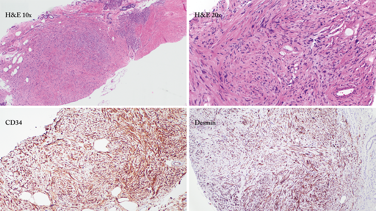
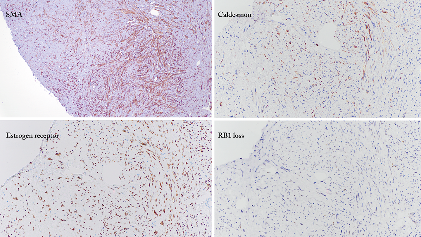
The following are the images from the second lesion.
Histology
What is your diagnosis?
a) Metaplastic carcinoma
b) Leiomyosarcoma
c) Myofibroblastoma with atypical leiomyomatous features
d) Fibromatosis
e) Solitary fibrous tumor
Click here to register your guess and see the answer.
Do you have an interesting case that you would like us to feature? Email it to edit@thepathologist.com.
Submitted by Mohan Narasimhamurthy, Assistant Professor of Pathology, Microbiology, and Immunology, and Melinda Sanders, Professor, Department of Pathology, Microbiology and Immunology, Vanderbilt University Medical Center, Nashville, Tennessee, USA.
Newsletters
Receive the latest pathologist news, personalities, education, and career development – weekly to your inbox.


