Conexiant
Login
  • The Analytical Scientist
  • The Cannabis Scientist
  • The Medicine Maker
  • The Ophthalmologist
  • The Pathologist
  • The Traditional Scientist
The Pathologist
  • Explore Pathology

    Explore

    • Latest
    • Insights
    • Case Studies
    • Opinion & Personal Narratives
    • Research & Innovations
    • Product Profiles

    Featured Topics

    • Molecular Pathology
    • Infectious Disease
    • Digital Pathology

    Issues

    • Latest Issue
    • Archive
  • Subspecialties
    • Oncology
    • Histology
    • Cytology
    • Hematology
    • Endocrinology
    • Neurology
    • Microbiology & Immunology
    • Forensics
    • Pathologists' Assistants
  • Training & Education

    Career Development

    • Professional Development
    • Career Pathways
    • Workforce Trends

    Educational Resources

    • Guidelines & Recommendations
    • App Notes

    Events

    • Webinars
    • Live Events
  • Events
    • Live Events
    • Webinars
  • Profiles & Community

    People & Profiles

    • Power List
    • Voices in the Community
    • Authors & Contributors
  • Multimedia
    • Video
    • Podcasts
    • Pathology Captures
Subscribe
Subscribe

False

The Pathologist / Issues / 2025 / June / Multimodal Deep Learning Model Sets New Benchmarks in Medical Imaging
Oncology Digital and computational pathology Bioinformatics Technology and innovation Software and hardware Clinical care Screening and monitoring Research and Innovations Microscopy and imaging

Multimodal Deep Learning Model Sets New Benchmarks in Medical Imaging

New framework integrates radiology and pathology imaging to support classification tasks in diagnostic imaging

06/26/2025 News 2 min read

Share

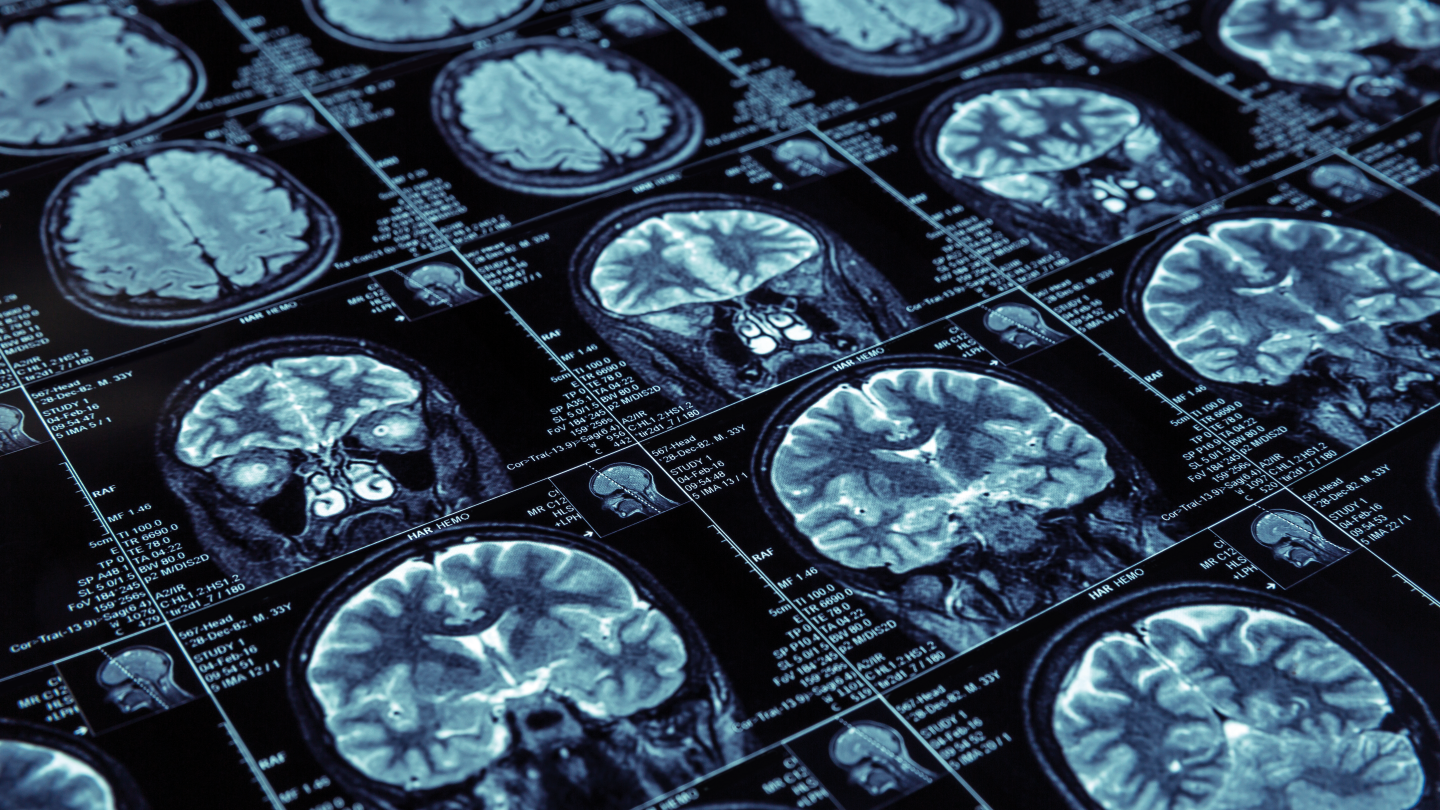
Credit: Adobe Stock

Researchers have developed a deep learning framework that integrates radiology and pathology imaging to support classification tasks in diagnostic imaging. The framework combines the Adaptive Multi-Resolution Imaging Network (AMRI-Net) with the Explainable Domain-Adaptive Learning (EDAL) strategy in a unified architecture. According to the study published in Frontiers in Medicine, this approach is designed to address the variability of multimodal medical data and improve the interpretability of artificial intelligence (AI) models used in image classification.

The framework was evaluated using four publicly available datasets: ISIC, HAM10000, OCT2017, and Brain MRI. Transformer-based backbone architectures and a cross-entropy loss function were used to train the classification model. AMRI-Net applies a multi-resolution feature extraction method with attention-guided fusion to process both local and global imaging patterns. EDAL introduces domain generalization techniques, including maximum mean discrepancy and adversarial domain alignment, and uses attention-based Grad-CAM to support model interpretability by highlighting regions associated with the model’s predictions.

In the ISIC dataset, which includes dermoscopic images, the framework achieved a classification accuracy of 95 percent and an F1 (precision and recall) score of 95 percent. These results by Lanting He of the School of Optoelectronics, Beijing Institute of Technology, and colleagues were higher than those obtained using baseline models such as BLIP (94 percent accuracy) and Vision Transformer (ViT). For the HAM10000 dataset, the framework reached an accuracy of 88 percent and an F1 score of 91 percent. On the OCT2017 dataset, the model achieved 89 percent accuracy, 96 percent recall, and an F1 score of 87 percent. For the Brain MRI dataset, the reported accuracy was 85 percent, with an F1 score of 86 percent.

An ablation analysis indicated that removing either AMRI-Net or EDAL from the framework resulted in lower classification metrics, suggesting each component contributed to overall performance. On the ISIC dataset, interpretability evaluations produced an Intersection over Union of 0.64 and pointing game accuracy of 81 percent, which were higher than the corresponding values from ResNet-CAM and ViT-based attention mechanisms.

The study findings indicate that the framework performs consistently across multiple datasets and includes interpretability features that may be applicable in diagnostic imaging settings with varying resource availability.

Newsletters

Receive the latest analytical scientist news, personalities, education, and career development – weekly to your inbox.

Newsletter Signup Image

Explore More in Analytical Science

Dive deeper into the analytical science. Explore the latest articles, case studies, expert insights, and groundbreaking research.

False

Advertisement

Recommended

False

Related Content

Global Referral
Digital and computational pathology
Global Referral

January 12, 2024

10 min read

How digital pathology is transforming the delivery of remote second opinions

Cracking Colon Cancer
Digital and computational pathology
Cracking Colon Cancer

January 25, 2024

1 min read

How a new clinically approved AI-based tool enables rapid microsatellite instability detection

The (Pathology) IT Crowd?
Digital and computational pathology
The (Pathology) IT Crowd?

December 30, 2021

5 min read

The pathologist’s guide to IT considerations for digitization

Defining the Next Generation of NGS
Digital and computational pathology
Defining the Next Generation of NGS

December 31, 2021

1 min read

Overcoming challenges of the typical NGS workflow with the Ion Torrent™ Genexus™ System

False

The Pathologist
Subscribe

About

  • About Us
  • Work at Conexiant Europe
  • Terms and Conditions
  • Privacy Policy
  • Advertise With Us
  • Contact Us

Copyright © 2025 Texere Publishing Limited (trading as Conexiant), with registered number 08113419 whose registered office is at Booths No. 1, Booths Park, Chelford Road, Knutsford, England, WA16 8GS.