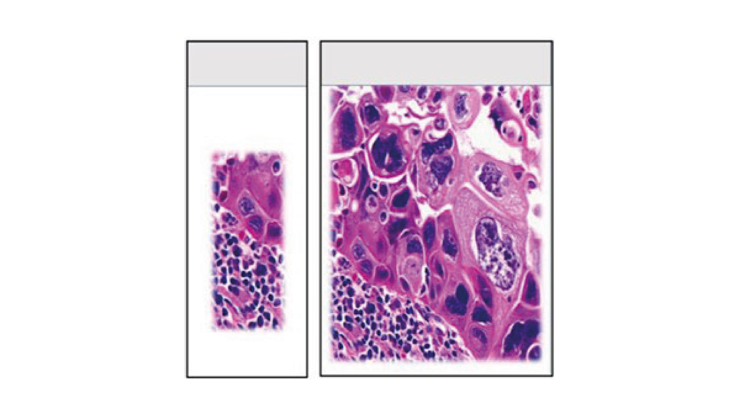
Whole mount histology (WMH) is a procedural system that is easily adapted to the needs of clinical and research laboratories. Also known as large format or supa mega histology, the technique is applied routinely to examine intact cross-sections of large tissue samples that are not usually achievable using the traditional small block sampling method.
Although the large format approach was first introduced more than a century ago to study breast cancers (1), it has in more recent times become a recognized method in the multidisciplinary approach to cancer diagnosis. In 2012, a special edition of the International Journal of Breast Cancer was influential in directing laboratories to introduce the large format system into routine diagnostic practice (2).
Around that time, an eight-year study aimed at contrasting the power of large format versus small block processing for breast cancer was completed (3). Surprisingly, results showed areas of histological significance in a high proportion of the large format slides. As these findings were absent on conventional slides, the results had the potential to modify clinical outcomes.

Going large
The routine practice of using small blocks often results in underreporting of the extent of diffuse malignancies by destroying the interrelationship of tumor components. In addition, evaluation of surgical margins is often unreliable as they are not always represented in the same block (4). By using WMH methods, these limitations are overcome as they are based on the processing of consecutive tissue slices that represent entire cross sections of tissue samples.
Criticisms that technical complexity impedes integration of the large format system into laboratory workflows are unjustified (5). The technique is easily adapted for use with existing laboratory equipment such as tissue processors and microtomes, and the stages for the processing of whole mounts are identical to those used in traditional methods.
Easily integrated alongside standard practices, WMH is regularly used throughout the United Kingdom and Europe and continues to gain global popularity. Consequently, laboratories are likely to meet new developments as companies strive to streamline their whole mount systems into standard practices.
Current developments include mega cassette printers, slide printers and label printers. There are also automated platforms such as the IntelliPath FLX that will accept double width slides for immunohistochemistry and in situ hybridization. In addition, for those fortunate enough to be equipped with imaging technology, digitized whole-slide images are possible with large slide scanners such as the Aperio, the NanoZoomer, and the TissueScope CF.
Power to the prognosis
In the diagnostic laboratory, the large format method allows correct identification of important prognostic factors such as tumor size, extent of disease, and the status of the circumferential surgical margin. Furthermore, detailed and systematic correlation studies of pathological and radiological scanning modes such as mammography, CT, and MRI together provide histological and spatial evidence that is invaluable in the multidisciplinary approach to cancer diagnosis (4).
With similarities between the morphological, spatial and technical requirements of breast and prostate cancers, large format histology has proven to be a powerful tool in the investigation of radical prostatectomy specimens. Because prostate cancers are not always obvious at gross examination, processing the entire prostate avoids under sampling of the tumor.
Although traditional small block sampling of the entire prostate is often adequate, WMH maintains the integrity of tissue sections during processing. Displaying the entire cross section allows for a greater appreciation of the organ architecture, and more clarity in identification of tumor nodules. It also facilitates direct comparison between histology and medical imaging findings (6). Furthermore, by enabling a higher detection rate of adverse pathology, better outcomes are provided for prostate cancer patients.
Accuracy in oncology
Large format histology is frequently applied to cancers of the breast and prostate yet has been shown to be a powerful tool in benign and neoplastic disorders of other organs. For example, WMH has been used to examine the pathological features of laryngectomy specimens in the diagnosis and management of laryngeal cancer.
Gross pathology and histomorphology of laryngeal specimens following surgery are the most accurate methods for determining the extent of tumor involvement. Consequently, the part of the larynx involved by the lesion should be entirely submitted for whole mount examination as it has been shown to be superior to standard slide sectioning of laryngectomy specimens (7).
Similarly, in radical nephrectomies, the system has proved invaluable in clarifying the relationship of renal tumors to the perinephric fat, which is not always apparent in small sized blocks (8). Since perinephric fat invasion is often subtle, accurate identification of it is often critical for staging cancers of the kidney.
Equally, in patients with rectal cancer, the crucial factors influencing the risk of local recurrence and poor survival are the status of the circumferential resection margin and quality of the mesorectal excision. Although whole mount sections often provide a more accurate assessment of rectal cancer that has infiltrated the mesorectum, comparison studies using both the large format and small block processing systems have shown similar results when resection margin status is assessed (9).
Bioengineering and beyond
Moving away from cancer diagnostics, the application of WMH has been effective in an emerging area of research that integrates tissue engineering and biological sciences with advanced manufacturing. By creating personalized healthcare solutions such as patient-specific implants for reconstructive surgery, biofabrication has been used in the treatment of microtia, a congenital condition which affects the formation of the external ear (10).
Another fascinating application of WMH is in the combined use of histology and MRI imaging to obtain high-resolution 3D images of the human brain (11). By using the large format method to minimize block numbers while preserving the integrity of subcortical structures, a high-resolution computational atlas of the human brain has been produced – based on 3D reconstructed histology.
The reconstruction of artificial whole mount sections from digitized tissue fragments is also possible using software tools (12). The algorithm used has the capacity to aid pathology workflows by bringing the benefits of large format systems to laboratories that are unable to employ them.
And lastly, an interesting veterinary application of the system: the study of Alzheimer’s disease in stranded dolphins, where presence of the neuropathological hallmarks of the disease (amyloid-beta plaques, phospho-tau accumulation, and gliosis) were diagnostic (13). Brain pathology helped to explain cases of live strandings and their ultimate deaths – supporting the “sick-leader” theory whereby healthy members in a pod mass become stranded as a result of high social cohesion.
Driving disease understanding
Various applications and advantages of large format histology in the research and clinical laboratory have already been described. In the diagnostic laboratory, the ability to provide improved sampling and preservation of the spatial arrangement of tissues is fundamental for studying the characteristics and heterogeneity of disease processes such as cancer.
Additionally, the correlation of pathological findings with those obtained from imaging studies, the development of new methods to produce anatomic and diagnostic libraries, and the integration of tissue engineering with biological sciences have provided whole mount histology with a very promising future.
References
- GL Cheatle, “Early recognition of cancer of the breast,” British Medical Journal, 1 (1906).
- T Tot et al., “Large format histology in diagnosing breast carcinoma,” Int J Breast Cancer, 2012 (2012). PMID: 23326672.
- M R Foster et al., “Large format histology may aid in the detection of unsuspected pathologic findings of potential clinical significance,” Int J Breast Cancer, 2012, (2012). PMID: 23050156.
- L Tabár et al., “Can we improve breast cancer management using an image-guided histopathology workup supported by larger histopathology sections?” Eur J Radiol, 161 (2023). PMID: 36821956.
- P Bryant et al., “Application of large format tissue processing in the histology laboratory,” J Histotechnol, 42, 3 (2019). PMID: 31492093.
- L Duan et al., “Advantage of whole-mount histopathology in prostate cancer: current applications and future prospects,” BMC Cancer, 24, 448 (2024). PMID: 38605339.
- M A Swid et al., “Operative techniques in otolaryngology: Head and neck surgery. Geisinger Medical Center experience for whole mount laryngectomy specimen,” Oper Tech Otolayngol Head Neck Surg, 35, 2 (2024).
- M Varma and J Dormer, “Macroscopy of specimens from the genitourinary system,” J Clin Pathol, 77, 3 (2024). PMID: 38373783.
- F Giner et al., “A comparison of whole-mount and conventional sections for pathological mesorectal extension and circumferential resection margin assessment after total meso-rectal excision,” Cir Esp (Engl Ed), 102, 8 (2024). PMID: 38373616.
- MT Ross . “Novel imaging and bioprinting approaches for auricular cartilage regeneration,” PhD thesis, Queensland University of Technology (2022). doi:10.5204/thesis.eprints.235047
- M Mancini et al., “A multimodal computational pipeline for 3D histology of the human brain,” Sci Rep, 10, 1 (2020). PMID: 32796937.
- D Schouten et al., “Full resolution reconstruction of whole‑mount sections from digitized individual tissue fragments,” Sci Rep, 14, 1 (2024). PMID: 38233535.
- M C Vacher et al., “Alzheimer’s disease-like neuropathology in three species of oceanic dolphin,” Eur J Neurosci, 57, 7 (2023). PMID: 36514861.


