Conexiant
Login
  • The Analytical Scientist
  • The Cannabis Scientist
  • The Medicine Maker
  • The Ophthalmologist
  • The Pathologist
  • The Traditional Scientist
The Pathologist
  • Explore Pathology

    Explore

    • Latest
    • Insights
    • Case Studies
    • Opinion & Personal Narratives
    • Research & Innovations
    • Product Profiles

    Featured Topics

    • Molecular Pathology
    • Infectious Disease
    • Digital Pathology

    Issues

    • Latest Issue
    • Archive
  • Subspecialties
    • Oncology
    • Histology
    • Cytology
    • Hematology
    • Endocrinology
    • Neurology
    • Microbiology & Immunology
    • Forensics
    • Pathologists' Assistants
  • Training & Education

    Career Development

    • Professional Development
    • Career Pathways
    • Workforce Trends

    Educational Resources

    • Guidelines & Recommendations
    • App Notes

    Events

    • Webinars
    • Live Events
  • Events
    • Live Events
    • Webinars
  • Profiles & Community

    People & Profiles

    • Power List
    • Voices in the Community
    • Authors & Contributors
  • Multimedia
    • Video
    • Podcasts
Subscribe
Subscribe

False

The Pathologist / Issues / 2025 / Jan / Multidimensional Precision
Technology and innovation Point of care testing

Multidimensional Precision

How smartphone-based microscopy opens up imaging at the point of care

By Jessica Allerton 01/30/2025 News 2 min read

Share

A new smartphone-based digital holographic microscope (DHM) allows users to make precise 3D measurements with a broad range of applications (1). Here, lead researcher Yuki Nagahama shares more about this exciting development.

How does your smartphone-based DHM device differ from traditional systems?

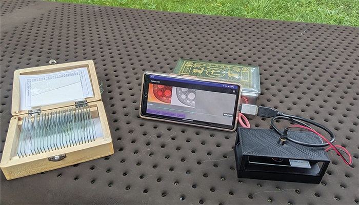
In this DHM system, laser light illuminates the object, creating interference fringes by combining light that passes through the object with light that does not. These fringes, called holograms, are recorded. A computational system then simulates light propagation on the hologram, allowing the object to be focused and observed later.

Unlike traditional microscopes, DHMs capture both the amplitude and phase information of light. This enables the observation of transparent objects, such as microorganisms and cells, without the need for staining.

How could this device enhance point-of-care (POC) testing, particularly in remote or resource-limited settings? 

We estimate this device will cost only tens of dollars in materials. With smartphones becoming more widespread, offering a microscope at this low price makes it far more affordable than typical options. This affordability could allow for the development of POC testing environments in a variety of locations.

What diagnostic advantages does this system offer for clinical pathology?

The phase information can be used to estimate the object's thickness, aiding in the identification of microscopic features.

Unlike typical microscopes, this DHM system doesn't use physical lenses, like eyepieces or objectives, to magnify images. Instead, it uses a touchscreen zoom function powered by optical simulation. This allows users to zoom in without changing lenses, enhancing convenience and usability.

How could the device be improved to broaden its applications in bio-imaging or enhance its value in clinical diagnostics?

We are exploring ways to enhance image quality with deep learning. DHMs often produce unwanted effects, like conjugated images, that overlap with the observed image. Our research focuses on using deep learning to remove these unwanted elements through image conversion.

More broadly, how do you anticipate POC technologies will evolve over the next decade?

With the growing demand for telemedicine, patients may increasingly perform tests at home or in remote areas and share results with medical professionals for diagnosis. Linking these devices with wearables and smart devices will also allow patients to monitor and manage their health. To enable this, a secure system for managing POC data and a robust communication infrastructure are essential. Additionally, providing support to help patients use POC devices effectively will be crucial.

Newsletters

Receive the latest pathology news, personalities, education, and career development – weekly to your inbox.

Newsletter Signup Image

References

  1. Y Nagahama, Optica, 63, 25 (2024). DOI: 10.1364/AO.532972.

About the Author(s)

Jessica Allerton

Deputy Editor, The Pathologist

More Articles by Jessica Allerton

Explore More in Pathology

Dive deeper into the world of pathology. Explore the latest articles, case studies, expert insights, and groundbreaking research.

False

Advertisement

Recommended

False

Related Content

Intense Intestines
Microscopy and imaging
Intense Intestines

January 19, 2024

1 min read

Some powerful fluorescent microscopy in this Image of the Month…

Context Matters in Cancer Biology
Microscopy and imaging
Context Matters in Cancer Biology

December 27, 2021

1 min read

Akoya is leading the way with spatial phenotypic signatures – a novel class of biomarkers for predicting response to immunotherapy

When Pathology Goes 3D
Microscopy and imaging
When Pathology Goes 3D

February 2, 2022

1 min read

Stratifying cancers with non-destructive 3D pathology

Sacrificing Safety for Speed?
Microscopy and imaging
Sacrificing Safety for Speed?

February 4, 2022

3 min read

Wide adaptation of antigen testing could increase the risk of exposure to infectious SARS-CoV-2 samples

False

The Pathologist
Subscribe

About

  • About Us
  • Work at Conexiant Europe
  • Terms and Conditions
  • Privacy Policy
  • Advertise With Us
  • Contact Us

Copyright © 2025 Texere Publishing Limited (trading as Conexiant), with registered number 08113419 whose registered office is at Booths No. 1, Booths Park, Chelford Road, Knutsford, England, WA16 8GS.