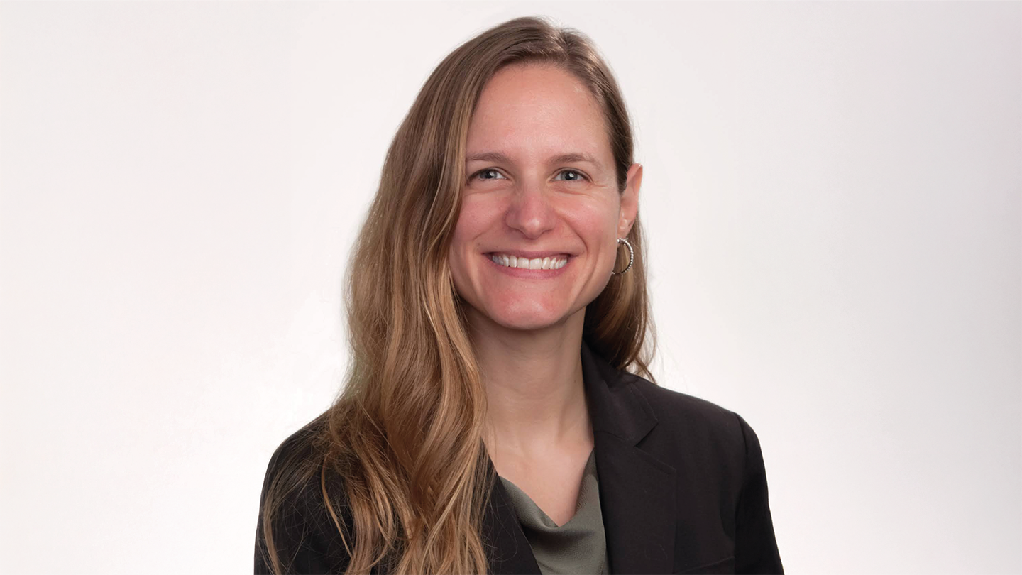
Meet Amanda Hemmerich
I am board certified in anatomic and clinical pathology. After completing residency training at Duke University, I moved to the University of North Carolina to do a specialized surgical pathology fellowship. I also gained specialized experience in gastrointestinal and liver pathology there.
I then returned to Duke University and did my cytopathology fellowship, which gave me insightful experience working with different providers in the room during procedures and resulted in another board-certification.
After a brief stint in private practice, I came back to Duke University and worked as an academic gastrointestinal and liver pathologist for about a year. But I felt that I was more suited for and interested in working in the life sciences industry, whether consulting or in another business role. I reached out to my contacts in academia to see what opportunities may be available to me.
I took a position with Foundation Medicine – a purely molecular company offering next-generation sequencing (NGS) testing of tumors. I liken it to throwing the kitchen sink at a lesion to identify what may be happening, why and how it's growing, and then pairing it with the approved therapeutics or even clinical trials, which may benefit that particular patient.
There was also a huge explosion in PD-L1 testing for checkpoint inhibitors while I was there, and I was involved in developing different immunohistochemistry (IHC) assays.
With that molecular experience under my belt, I transitioned to IQVIA Laboratories and joined its molecular hub in Research Triangle Park, North Carolina. While at this innovation lab, I assisted with research in exploratory biomarker development that largely looked at the expression of proteins of interest via IHC assays.
IQVIA is a large, global clinical research organization; IQVIA Laboratories is the clinical lab services arm. We can develop new assays for exploratory trials or run proprietary tests for retrospective studies. Along with managing the regulatory documentation and audit trails for the testing, we also work closely with our clients to establish the optimum data output – right down to the scanning parameters and image file format.
How did you get started in computational pathology?
John Cochran, Chief Medical Officer and Chief Pathologist at IQVIA Laboratories, recognized that my skills would translate well to oversight of the digital pathology initiative. And I was up for the challenge!
First, I learned all about the various scanning technologies, file formats, image management systems, and data storage options for the research space. Next, I looked into image analysis, exploring the answers to key questions. How does machine learning happen? What does deep learning and artificial intelligence do? What’s a foundational model? How many different image analysis vendors are there?
I needed to navigate how our IT system would deal with digital pathology, what the audit trail would look like, and how to train our personnel to work with it. It has also given me the chance to collaborate internationally with our labs across the world, in Scotland, the US, Singapore, and Beijing.
Setting up the digital pathology system has been a huge (and exciting) learning curve. It has also been a lot of fun.
How is clinical research harnessing image analysis software?
AI introduces a useful tool for pattern recognition, but it has taken a long time to get it right. Imagine training an algorithm to recognize a dog. It would learn from a set of 100 dog images. But what if you challenged it with an image of a cat. The program sees two ears, four legs, a nose and a tail and recognizes it as a dog. Now, with newer foundational models, the training starts with images of normal, healthy tissue. So, when it’s shown cancerous tissue, the model can recognize it as abnormal tissue.
Currently, we’re at the stage where we can start using these tools for image analysis. Indeed, trained algorithms can give highly accurate percentages of stained tumor cells on IHC slides or calculate ratios like cytoplasm to membranous staining.
We’re also seeing the emergence of companion diagnostic algorithms, which are allowing us to analyze tissue sample images from patients on specific drugs and calculate cell ratios to see if the patient is actually benefiting from the treatment. It is simply not feasible for a pathologist to come up with that sort of scoring system just from looking at a glass slide, which makes these tech-enabled solutions vital supplements to our expert eyes.
Hopefully, in the future, we will have image analysis algorithms that predict the most likely alteration in a specific type of tumor, so we know where to focus our testing.
Not only are these sorts of targeted approaches highly efficient, but they are also helping to deliver more personalized patient care, which is ultimately the goal.
How is digital pathology and AI enhancing clinical trial efficiency and access?
There is still work to be done to fully realize the potential of digital pathology in research. Ideally, we need all the local hospitals who participate in trials to have access to slide scanners. And, if we could standardize the file formats of digital images, it would help a lot. Even if hospitals have to send slides to a centralized lab for scanning, the resulting digital whole-slide images allow trials to be location agnostic, which really helps with efficiency and workflows. For example, we could have our pathologist in Mumbai, India, look at a case so that when the patient in the UK wakes up, their results are ready and waiting for them, confirming whether they can start the clinical trial that day. In that way, pathology becomes much more of a 24/7 operation.
There are other practical benefits when you have pathologist specialists spread across various locations around the world. For example, if our hematopathologist in the UK was unavailable but we needed someone else to look at the cases, we would have had to package the glass slides up and ship them to California, adding weeks to the timeline. With digitized slides, the diagnostic workflow is no longer hampered by delays or complications due to shipping.
Something as simple as annotation tools on image management systems also make a huge difference to collaborative working. Pathologists in different locations, who are looking at the same digital slide, can now share notes and second opinions on a case as if they are in the same room.
Another aspect is quality control. We have AI tools that can assess the quality of staining on slides, so we can adjust accordingly to achieve more consistency. There are also algorithms that can assess the diagnostic accuracy of the pathologists themselves and spot any trends that emerge, which helps us plan training.
It’s crucial that pathologists are involved in clinical trials because they can advise on any discrepancies between the assays in the trial environment and how they are likely to be performed in the real world. For instance, if a particular scoring system is used in the study, it’s important to make sure it’s reproducible in a regular lab in an everyday clinical setting.
What innovations do you predict will emerge in the computational pathology space?
At the moment, I think that innovation is being hampered by proprietary image file formats, which results in incompatibilities or makes image sharing harder. Similarly, some companion diagnostics can only be used with vendor-specific slide scanners and image analysis tools, which makes end-to-end solutions cost prohibitive for a lot of labs.
I expect that the regulators may start to insist on standardized file formats. Hopefully, it will be the DICOM standard, which has been proven to aid interoperability in radiology.
I also predict more capitalization of IHC image analysis to develop companion diagnostics in oncology and beyond in the next two or three years.


