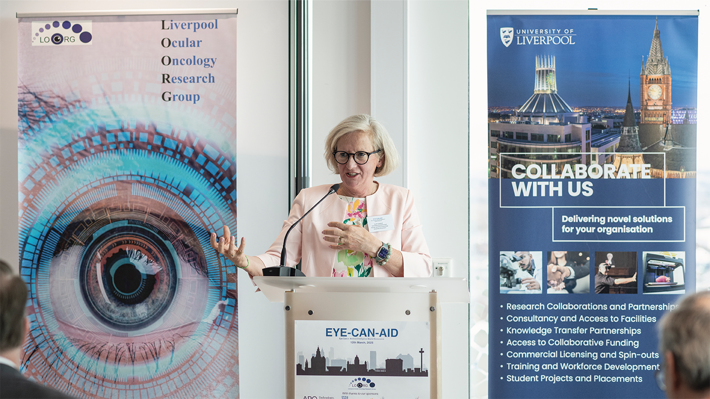
Researchers at the University of Liverpool have launched an initiative to advance our understanding of cancers of the eye. The Eye Cancer Artificial Intelligence Digital (EYE-CAN-AID) Bioresource, combines a digital eye cancer biobank with AI-driven analysis. It is hoped the resource will help to develop better diagnostic tools to inform personalized treatments.
Here, Sarah Coupland, Director of the Liverpool Ocular Oncology Research Group, reveals the technologies behind EYE-CAN-AID and what it could mean for patients with ocular cancer.
What is the EYE-CAN-AID initiative and how will it help to further our understanding of the underlying causes of ocular cancer?
The EYE-CAN-AID Bioresource initiative is a further development of the Liverpool Ocular Oncology Biobank, which has existed for 14 years. The Biobank contains tissue and matching blood samples from consented eye cancer patients linked with their clinical and genetic data – already an achievement in itself. EYE-CAN-AID incorporates these data with the respective digitized histology as well as ocular and radiological images.
EYE-CAN-AID offers a multimodal digital bioresource, which can allow for the detailed assessment of eye cancers at all levels and at different stages of the disease. Furthermore, by applying artificial intelligence to these images, it is hoped that they will reveal novel information that can be used to diagnose these tumors earlier and improve patient outcomes.
How does the addition of targeted slide scanner technology enhance the research initiative?
Our new slide scanner, from Roche Diagnostics, has automatic start and continuous loading of up to 40 trays. It enables EYE-CAN-AID to perform high volume, high-quality scanning of all the histology slides of the ~4000 eye cancer cases that are housed within the current Liverpool Ocular Oncology Biobank.
The number of histology slides varies per case but typically includes five of the main tumor block, including hematoxylin and eosin and some immunohistochemistry stains. The automated scanning will increase the speed at which all slides and all cases can be uploaded and stored within the trusted research environment of EYE-CAN-AID.
How is artificial intelligence being applied within EYE-CAN-AID?
Artificial intelligence has already been tested in the research arena, whereby it looks at whether AI can aid the judgment in differentiating between a “freckle” at the back of the eye versus a small cancer. Initial results on Liverpool data indicate that it can do this with high accuracy ; however, to validate this model further, we are extending to images collated at Moorfields Eye Hospital.
Another algorithm is being developed to differentiate between a choroidal melanoma versus a cancer that metastasizes to the choroid from a breast or lung cancer, for example. It should be noted that AI will not replace current technologies or indeed clinical acumen; it will just be an adjunct in the armory, providing further evidence when making diagnoses.
How has the work of the Liverpool Ocular Oncology Centre helped personalize treatment plans for eye cancer patients?
Eye cancers are rare, and so, if their data are disparate and not collated into one database, the evaluation of parameters relevant for clinical care will always suffer from inadequate statistical power and almost be anecdotal.
The Liverpool Ocular Oncology Centre commenced its database 32 years ago, when the center was established by the National Health Service in England. This database has already enabled the development of a robust stratification model of uveal melanoma patients that defines risk groups with respect to the development of metastases, typically occurring in the liver.
Accordingly, those patients considered to be high risk undergo more frequent liver surveillance using high resolution imaging. The aim is to detect liver metastases early enough to either surgically remove them or enlist the patient in a clinical trial. Those patients who are at low risk of developing metastases undergo less frequent liver surveillance, decreasing the anxiety associated with each scan and their related costs.
What are the anticipated long-term benefits of EYE-CAN-AID for patients?
EYE-CAN-AID collates all the ocular images of eye cancer patients into one trusted research environment, linked together with clinical treatments and outcomes, genetics and histology of the tumors. This will allow for their detailed analyses with respect to a range of factors, including the tumor size and location with the eye, and how this influences therapy choice, treatment dose, treatment success, and side effects. It will also enable predictions of which patient group is most likely to respond best to certain treatments, including novel ones within clinical trials.
Similar analyses could be undertaken in eye cancer patients who develop liver metastases. For example, are there particular sizes and locations of metastases that better respond to therapies, and if so, which ones?
What’s next in the evolution of EYE-CAN-AID?
We would like to extend our EYE-CAN-AID Bioresource to include the consented data from adult eye cancer patients treated in other eye cancer centers in the UK – namely Glasgow, London, and Sheffield. This would increase the data cohort and make the analyses even more robust.
We would also like to fully sequence the cancers arising in the eye, their respective metastases, and associated blood samples. The aim is to determine whether circulating tumor cells can be detected more reliably than present, permitting neoadjuvant therapies to be administered whilst metastatic tumor volume is low.
These plans are in motion, but will require infrastructural support and project management. Funding is being sought!


