Conexiant
Login
  • The Analytical Scientist
  • The Cannabis Scientist
  • The Medicine Maker
  • The Ophthalmologist
  • The Pathologist
  • The Traditional Scientist
The Pathologist
  • Explore Pathology

    Explore

    • Latest
    • Insights
    • Case Studies
    • Opinion & Personal Narratives
    • Research & Innovations
    • Product Profiles

    Featured Topics

    • Molecular Pathology
    • Infectious Disease
    • Digital Pathology

    Issues

    • Latest Issue
    • Archive
  • Subspecialties
    • Oncology
    • Histology
    • Cytology
    • Hematology
    • Endocrinology
    • Neurology
    • Microbiology & Immunology
    • Forensics
    • Pathologists' Assistants
  • Training & Education

    Career Development

    • Professional Development
    • Career Pathways
    • Workforce Trends

    Educational Resources

    • Guidelines & Recommendations
    • App Notes

    Events

    • Webinars
    • Live Events
  • Events
    • Live Events
    • Webinars
  • Profiles & Community

    People & Profiles

    • Power List
    • Voices in the Community
    • Authors & Contributors
  • Multimedia
    • Video
    • Podcasts
Subscribe
Subscribe

False

The Pathologist / Issues / 2023 / Apr / Pathology on Canvas
Hematology Profession Hematology Microbiology and Immunology Professional Development

Pathology on Canvas

A curation of reader artwork on a single, uniting subject: Pathology.

By Bibiana Campos Seijo, George Francis Lee, Georgia Hulme 04/10/2023 Gallery 4 min read

Share

Can you believe it’s been eight years since we first asked the pathology and laboratory medicine community to contribute to our inaugural image issue? And yet, in our octennial year, we’ve never had more creative, eye-catching, and surprising submissions to sift through.

For 2023, we are proud to feature a plethora of pieces in a variety of mediums – from abstract canvases dripping with H&E splendor to hand drawn illustrations that show off the sensorial experiences of grossing.

We are always impressed by the amazing talent that resides within pathology and laboratory medicine. But know that this gallery is but only a small snapshot – a single brushstroke on the giant canvas of this community’s skill and creativity.
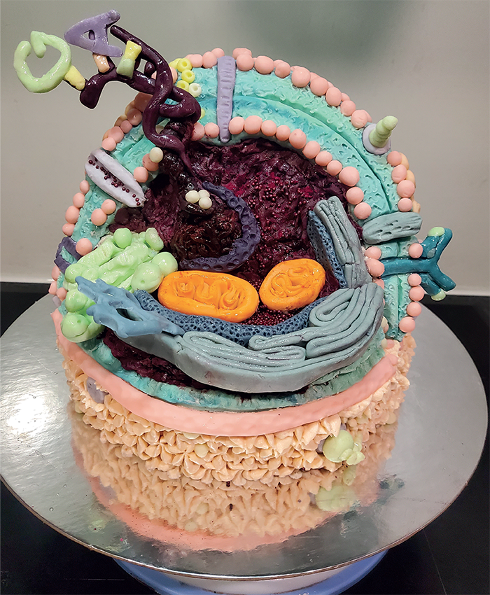
Inside a Cell

Credit: Alivea Smith-Andrews, Medical Laboratory Scientist, LabPLUS, Auckland City Hospital, Auckland, New Zealand
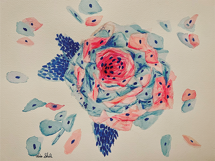
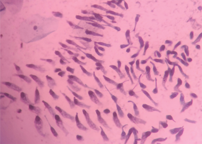
I made a cake depicting a B-cell's structure with organelles, antibodies, and major histocompatibility complex (MHC) class II proteins. It shows the nucleus containing chromatin, which spirals into a DNA helix with histones and shows individual bases. Most of the decorations are made of modeling chocolate; the cell membrane is buttercream.

Pap Rose Painting

Credit: Andrea Shields, PGY-2, Loma Linda University, Loma Linda, California, USA

I painted this picture after a cytology-GYN rotation because the squamous cells reminded me of petals.

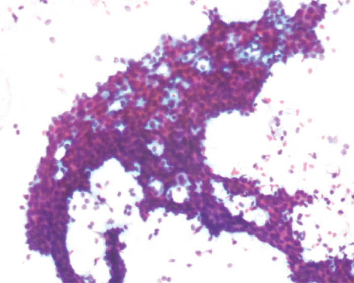
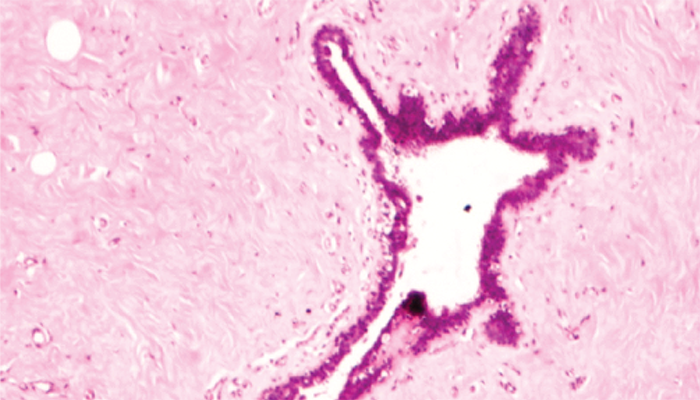
A Zoological Safari Through Tissue Specimens

Credit: Anoosha Murthy, Consultant Pathologist and Quality Manager, Celara Diagnostics, Bangalore, India.

Ductal Chameleon

Ductal Giraffe

Endocervical School of Fish

Ductal Crocodile Swallowing a Fish

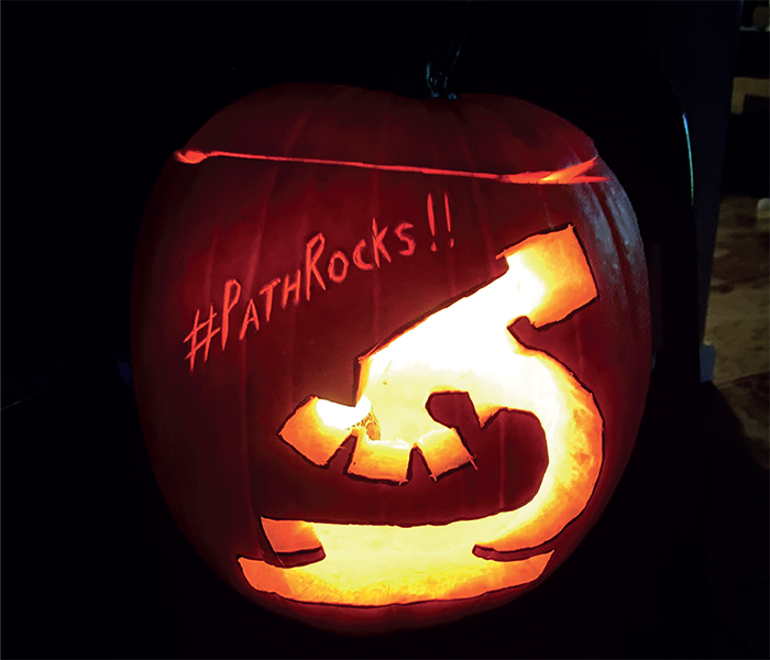
Halloween Carving, A Benign Abstract, Perched on a Tree

Credit: Apeksha Agarwal, Cytopathology Fellow, UT Health San Antonio, Texas, USA

Halloween Carving: I couldn't think of a better way to express my love for both pathology and Halloween.

A Benign Abstract: I was previewing a Pap stain of a benign thyroid nodule when its beautiful colors caught my attention.

Perched on a Tree: Cervical Pap smear resembles bird.

Perched on a Tree: Cervical Pap smear resembles bird.

Adipose Stems, Squamous Rainbow, The Wall Art

Credit: Aswathy Miriam Cheriyan, MD MPH, PGY 4 Anatomic & Clinical Pathology, Allegheny General Hospital, Pittsburgh, PA, US.

Adipose stems: Acrylic abstract painting on a 12x 12 in. canvas

Squamous Rainbow: Acrylic abstract painting on a 10x 10 in. canvas

The Wall: Acrylic abstract painting on a 10x 10 in. canvas

Perfect Flower

Credit: Chitturi Ramya, Associate Professor, NRI Medical College, Chinakakani, India

Fruiting body of Aspergillus fungus identified in the maxillary sinus tissue of a post-COVID-19 patient.

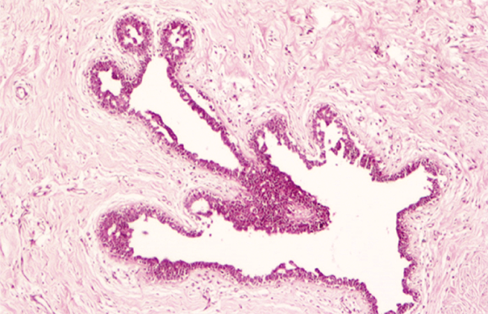
Sea of Cartilage, Trees of Ferning, Geometry of Colloid, Balloons and Vegetables, Flowers of Prostate

Credit: Evanthia Omoscharka, Pathology Residency Program Director and Associate Professor of Pathology, University of Missouri-Kansas City School of Medicine; Cytopathology and Point of Care Director, University Health, Kansas City, Missouri, US

Flowers of prostate: Corpora amylacea, 20 x magnification

Sea of cartilage: Cartilage, 40x magnification

Trees of ferning: KOH ferning, 20x magnification

Geometry of colloid: Colloid, 20x magnification

Balloons and vegetables: Vegetable material, 40x magnification

Little Bird, Gouty Stained Glass, Lymphoma Flowers

Credit: Faye Smith-Chakmakova, Pathologist, Barton Memorial Hospital, South Lake Tahoe, California, US

Little Bird: The bird is molluscum contagiosum in a biopsy of a penile lesion

Gouty Stained Glass: The stained glass is a gouty tophus under polarized light

Lymphoma Flowers: The flowers are high grade lymphoma in a pleural fluid

Stepping Stains to Histology

Credit: Frazer Bell, Histopathology Technician, Histology Research Service/ Veterinary Diagnostics Services, College of Medical, Veterinary and Life Sciences, University of Glasgow

Mouse embryo paws stained with H&E and Alcian blue/Masson’s trichrome.

Searching For a Cure

Credit: Jaye H. Paulman, Laboratory Manager at Southview Medical Group, Birmingham, AL, USA

This image was rendered in Daz Studio software using empiric assets with a laboratory setting. A touch-up for additional lighting was performed in Adobe Photoshop.

Chirp, Chirp

Credit: Linlin Gao, Department of Pathology and Laboratory Medicine, University of Kansas Medical Center, Kansas City, Kansas, USA

This image shows H&E staining with a group of red blood cells aggregated in the shape of a bird.

Fibroadenoma Fox

Credit: Lynn Messersmith, Pathologist, Martin Army Community Hospital, Ft. Benning, Georgia

Ductal elements create a curious fox in this section from a fibroadenoma resection. Hematoxylin & eosin at 40X magnification.

Pancreatic Tumor, Giant Cell Tumor of Bone, Glioblastoma

Credit: Mariana Duarte Riberio, Anatomical Pathology Unit, Coimbra Hospital and University Centre, Portugal.

Pancreatic Tumor: Concept art of a pancreatic tumor, located at the head of the pancreas with D2 compression. Knowledge of the anatomic relations is fundamental for understanding clinical symptoms as well as gross examination and sampling

Giant Cell Tumor of Bone: Concept art of a giant cell tumor (GCT) of bone. GCT is one of the most common bone tumors and it is easily recognized by its spatial heterogeneity. GCT induces marked bone destruction and may exhibit soft tissue extension. Based on an original image from the World Health Organization.

Glioblastoma: Concept art of a glioblastoma. Based on an original image from Pathorama (pathorama.ch).

Embroidered Kidney, Bone Brontosaurus

Credit: Meagan Chambers, Resident, Department of Pathology and Laboratory Medicine, University of Washington, Seattle, Washington, USA.

Embroidered Kidney: A fiber art creation to showcase this wonderful organ

Bone Brontosaurus: A dinosaur captured under the microscope

The Patient Behind the Lens

Credit: Michele Mitchell, Co-Chair of the University of Michigan Dept of Pathology Patient and Family Advisory Council

Michele Mitchell is a breast cancer survivor. Since retirement, she has devoted her time to patient advocacy. Michele takes the advocacy role seriously: “it is a great honor to educate and empower patients, move the needle on important issues, and make a real difference in healthcare policy, quality, and safety.”

Blood Cells, Breathe, Renal, Whipple, Thyroid and Trachea

Credit: Rachel Dunlap BA, MS, PA(ASCP)cm; Pathologists’ Assistant; Chicago Area Autopsy Service; Chicago, IL

Blood Cells: Watercolor on cotton paper. Recreation of a piece by LyonRoadArt.

Breathe: Abstract watercolor on cotton paper. Recreation of a piece by Katharine Asher.

Renal: Kidney watercolor on cotton paper.

Whipple: Pancreaticoduodenectomy with tail and gallbladder. Watercolor on cotton paper.

Thyroid and Trachea: Watercolor on cotton paper. Recreation of a piece by an unknown artist.

Pathology on Canvas

Credit: Regina Zavodovskaya, Associate Veterinary Anatomic Pathologist, VDx, Davis, California, USA.
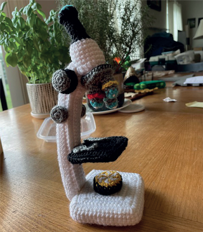
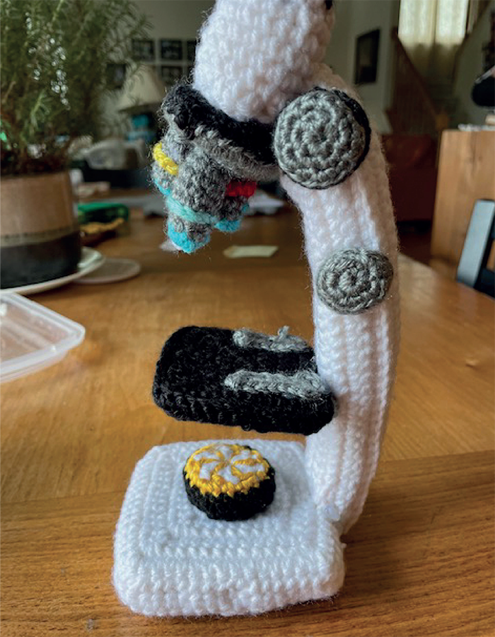
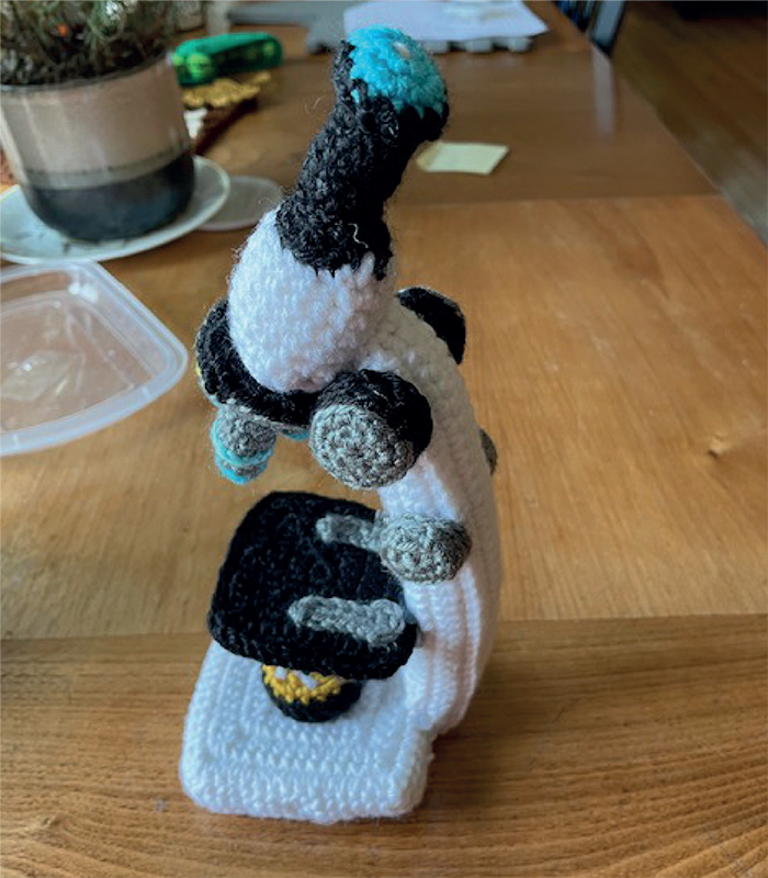
In this art piece, H&E stain colors were used as the primary theme.

Crochet Microscope

Credit: Renee Fraser, Cytotechnologist, Northwestern Medicine-West Region, Winfield, Illinois, USA

What do you give the lab professional who has everything?

I Love You with My Microscopic Hearts, Speed Doesn't Matter, My Fur Baby

Credit: Rico P. Lasaca, Department of Pathology, Divine Word Hospital, Tacloban, Philippines

I Love You with My Microscopic Hearts: A personal collection of images from different tissues and organs

Speed Doesn't Matter: An orange turtle discovered under the microscope

My Fur Baby: A canine friend in histology

Blood Cells, GI Mucosa, LSIL

Credit: Shabnam Seydafkan, Resident Physician, PGY-4, Department of Pathology, SUNY Downstate Health, Brooklyn, New York

Blood Cells: Acrylic painting

GI Mucosa: Acrylic painting

LSIL: Acrylic painting

The Brook in the Epidermal Cyst

Credit: Syed Salahuddin Ahmed, Senior Consultant in Pathology, Delta Hospital, Dhaka, Bangladesh

Photomicrograph of an epidermal cyst. Cyst content retracted from the wall gives the appearance of a brook.

A Pathologist’s Universe Is in the Microscope, Butterflies in a Glandular Garden

Credit: Vasudev Prabhu, Consultant Pathologist, Tejasvini Hospital, Mangaluru, India

A Pathologist’s Universe Is in the Microscope: In a universe of innumerable pathological conditions, the pathologist's tool, the microscope, is like a telescope – a powerful tool to explore a different universe of its own kind.

Butterflies in a glandular garden

Newsletters

Receive the latest pathology news, personalities, education, and career development – weekly to your inbox.

Newsletter Signup Image

About the Author(s)

Bibiana Campos Seijo

More Articles by Bibiana Campos Seijo

George Francis Lee

Interested in how disease interacts with our world. Writing stories covering subjects like politics, society, and climate change.

More Articles by George Francis Lee

Georgia Hulme

Associate Editor for the Pathologist

More Articles by Georgia Hulme

Explore More in Pathology

Dive deeper into the world of pathology. Explore the latest articles, case studies, expert insights, and groundbreaking research.

False

Advertisement

Recommended

False

Related Content

Real-Life Forensic Pathology Is Not CSI
Profession
Real-Life Forensic Pathology Is Not CSI

January 30, 2024

5 min read

Sitting Down With… Ken Obenson, Forensic Pathologist at The Saint John Regional Hospital, New Brunswick, Canada

Byte the Bias
Profession
Byte the Bias

January 11, 2024

1 min read

How does artificial intelligence view a pathologist?

Life Balance
Profession
Life Balance

January 5, 2024

9 min read

Sitting Down With… Kamran Mirza, Professor of Pathology and Director of the Division of Education Programs, Michigan Medicine, University of Michigan, Ann Arbor, Michigan, United States

Oldie, But a Goldie
Profession
Oldie, But a Goldie

January 2, 2024

1 min read

A shining specimen of microscopy in this Image of the Month

False

The Pathologist
Subscribe

About

  • About Us
  • Work at Conexiant Europe
  • Terms and Conditions
  • Privacy Policy
  • Advertise With Us
  • Contact Us

Copyright © 2025 Texere Publishing Limited (trading as Conexiant), with registered number 08113419 whose registered office is at Booths No. 1, Booths Park, Chelford Road, Knutsford, England, WA16 8GS.