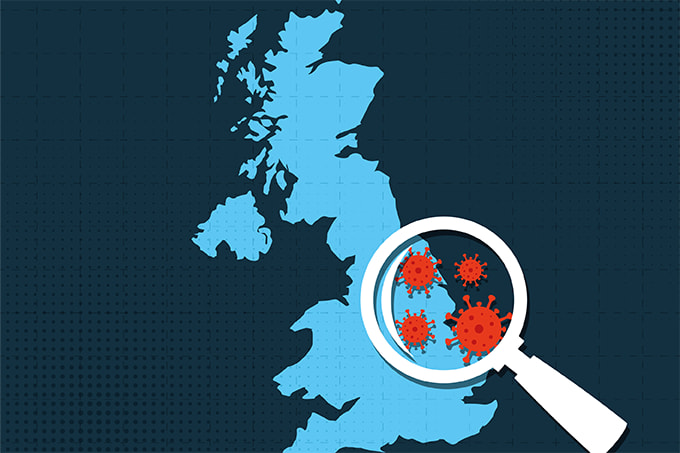
Two years into the pandemic, COVID-19 still presents a challenge for pathologists and researchers seeking to better understand how the disease affects the human body. Whole-slide imaging is central to this work because high-resolution images allow researchers to visualize and evaluate damage to tissues and cells. Though whole-slide images can be captured manually, the high volume of images needed for research requires a faster approach – so many researchers are turning to automated slide scanners to speed up the process and obtain higher-quality images. For example, Si Wang and colleagues used an automated slide scanner to study the lungs of patients with COVID-19 (1), whereas Ni Huang and others did the same to research COVID-19 infection of the oral cavity and saliva (2).
Automated slide scanners include many features to help researchers study infectious diseases – and a better understanding of COVID-19 means better patient outcomes. Automated slide scanners play an important role in disease research by providing fast, high-resolution imaging for quantitative analysis and publication. Here are six key scanner features I’ve found helpful in my own work.

1. Automated detection of samples during the scanning process.
Scanning whole slides of tissue sections and other samples for COVID-19 research can be time-consuming if the software scans both sample and background; automated sample detection speeds up the process by only detecting and scanning the sample. With accurate autofocus and high magnification on just the sample area, researchers can obtain all the information they need from the image while saving time scanning.
2. Intuitive software for an easy scan setup.
Easy-to-use scanner software makes it simple for researchers of all experience levels to obtain high-quality virtual slide images of tissues and cells. For example, software that saves project settings for specific samples enables researchers to start a quick batch scan with minimal supervision. This greatly reduces setup time and increases efficiency, especially for fluorescence imaging – a method often used in COVID-19 research to label SARS-CoV-2 RNA.
3. Accurate color reproduction of tissues and stains.
In COVID-19 research and other pathology applications, accurate color reproduction is vital to view stained samples as they are seen with the naked eye. Various technologies – such as modern, energy-efficient LEDs that can match the spectral characteristics of a halogen lamp – support accurate color reproduction. Furthermore, color-corrected cameras and ICC profiles facilitate accurate color and intensity reproduction on computer monitors. These features enable the purple, cyan, and pink stains often used in COVID-19 research to show up correctly in whole-slide images.
4. Automated immersion oil dispensing.
High-resolution images let researchers see small details, such as viruses. Scanning slides with immersion oil can provide high-resolution images to help researchers visualize infections; however, this process is typically done manually and can be time-consuming. An automated oil dispenser integrated into the scanner software can add the correct amount of oil to the scan area and remember the slides that have already been scanned with oil. This automated feature gives oil scanning the same convenience as using a dry objective.
5. Flexible imaging to visualize infections.
Depending on the sample and application, COVID-19 researchers may need a range of observation methods to visualize infections or stains. Scanners with multiple imaging modes enable researchers to clearly see different details of the sample, such as tissue or RNA. Having the option to mix and match methods in a batch scan offers even greater flexibility and efficiency, because researchers can process multiple sample types in one scan.
6. Links to image management systems.
COVID-19 research requires a large number of high-resolution images to understand how the virus affects the human body. Multiple images are stitched together into one large, tiled image to enable easy visualization of tissues or cells. Without an easy way to store and view large files, image management can become cumbersome and disorganized, but automated slide scanners that link to an image management system can enable image sharing and collaboration via a simple web viewer. This feature eliminates the need to transfer large images while retaining the ability to view high-resolution data.
Newsletters
Receive the latest pathologist news, personalities, education, and career development – weekly to your inbox.

References
- S Wang et al., “A single-cell transcriptomic landscape of the lungs of patients with COVID-19,” Nat Cell Biol, 23, 1314 (2021). PMID: 34876692.
- N Huang et al., “SARS-CoV-2 infection of the oral cavity and saliva,” Nat Med, 27, 892 (2021). PMID: 33767405.