You show us the world of pathology through your eyes with a collection of the most beautiful, informative and intriguing images from every field of pathology and laboratory medicine. In this gallery, a single slide is worth a thousand words…
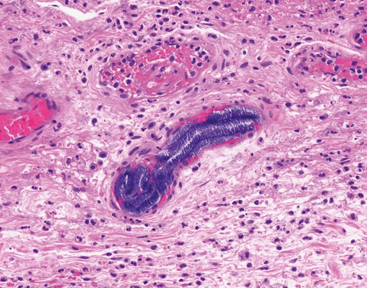
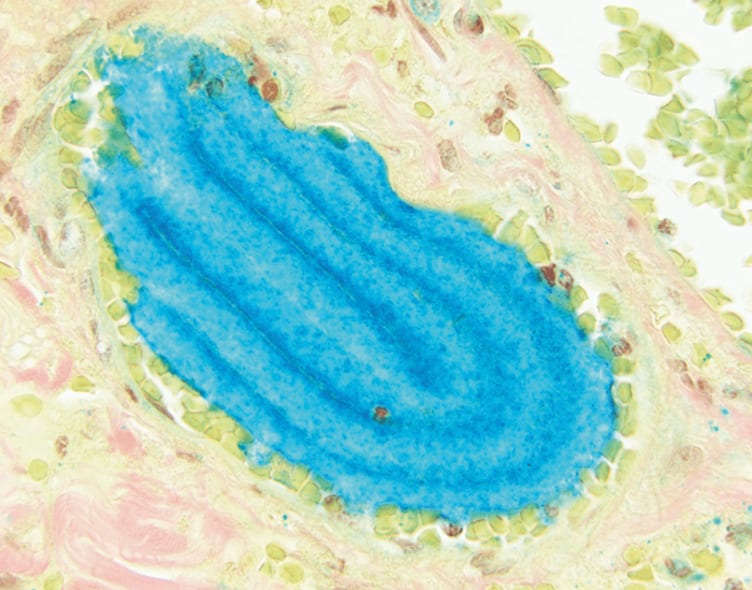
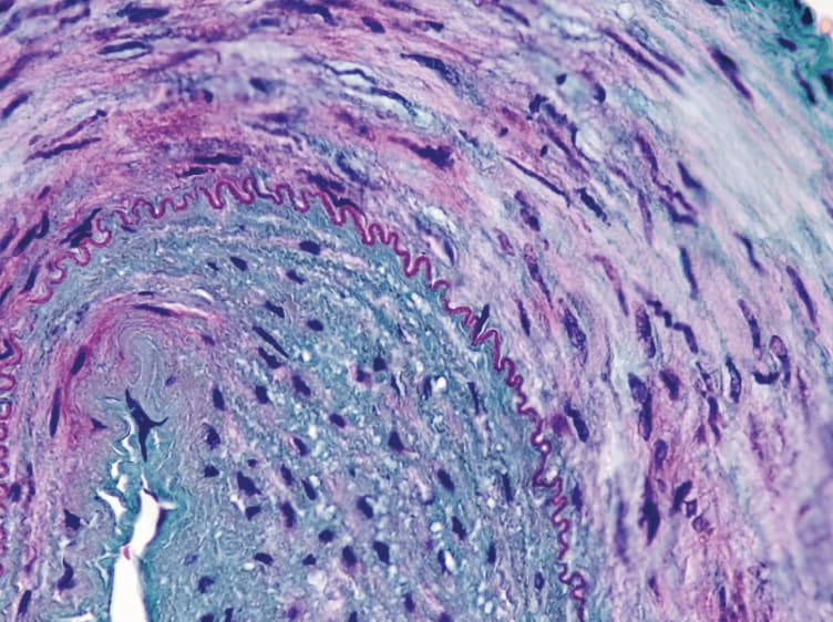
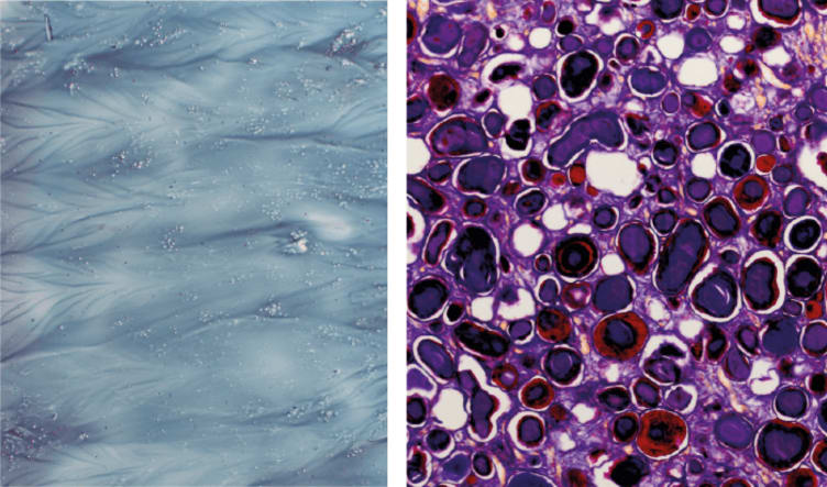
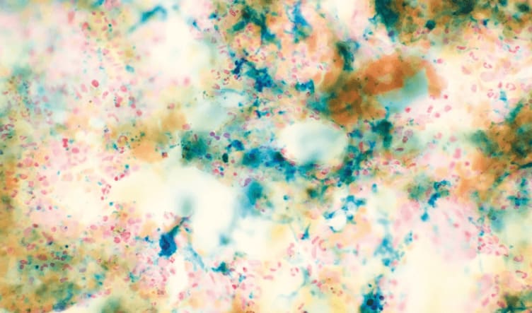
Hydrophilic Polymers
Hydrophilic polymers identified from a case of ischemic colitis in a patient with an aortic aneurysm repair. The polymers display a serpiginous configuration within vessels and a fuzzy and stippled basophilia on H&E (top) and are turquoise on colloidal iron (bottom).
Christina Arnold, The Ohio State University, USA.
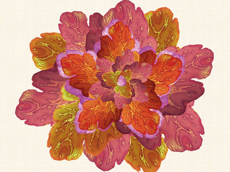
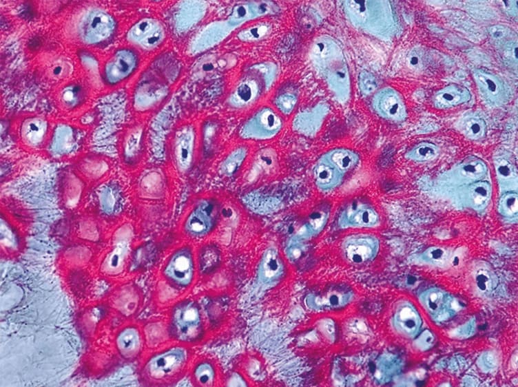
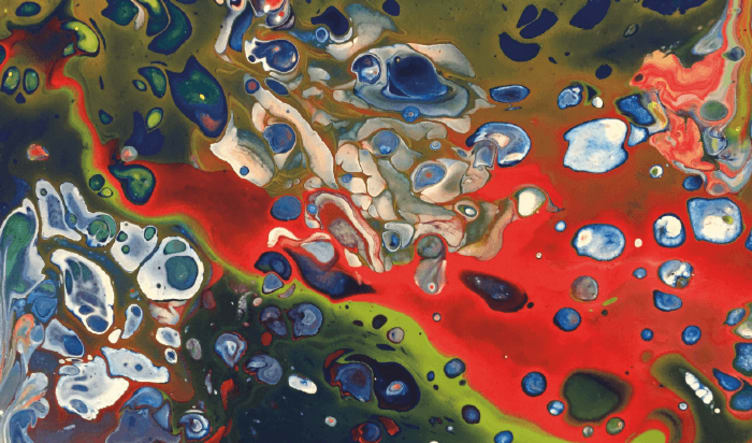
Blooming Condyloma
This piece was generated by Adobe Photoshop from a condyloma acuminatum, or genital wart.
Maha Abdulla, The University of Kanasas School of Medicine, USA.
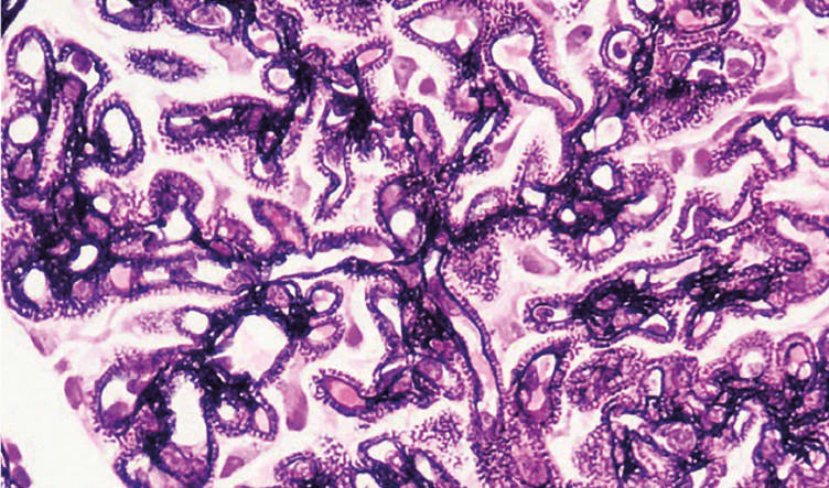
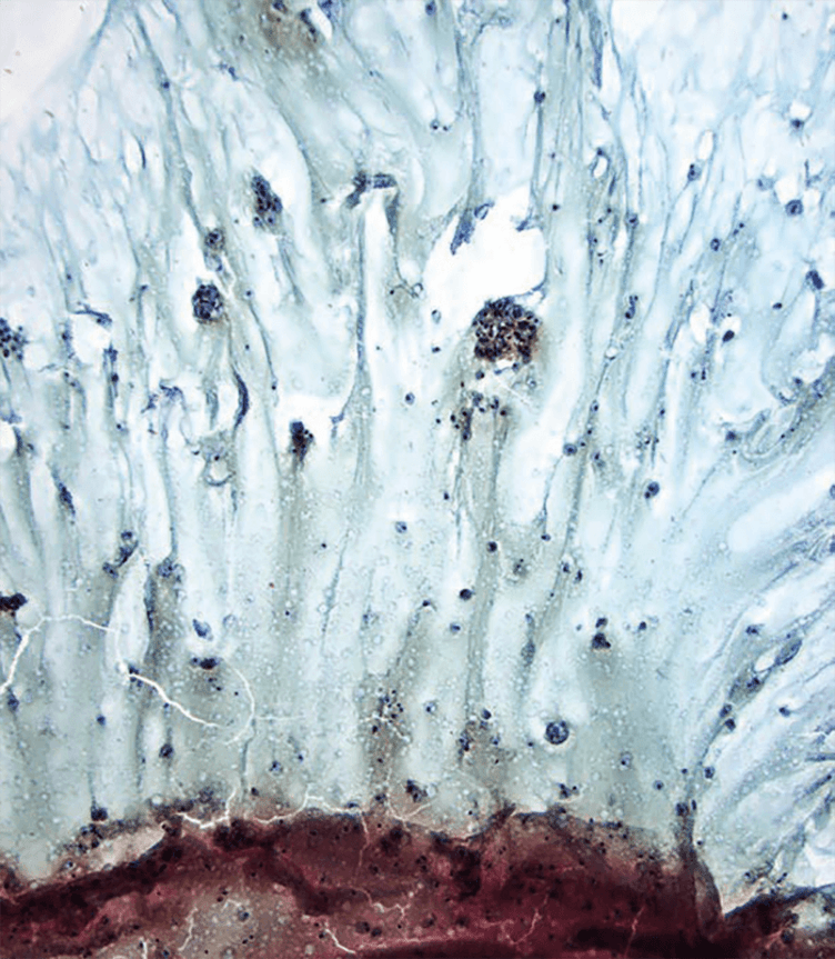
Elastic Cartilage
These images of elastic cartilage are stained by H&E and Nejib’s stain.
Luis Humberto Cruz, Mother and Child Hospital, Irapuato, Mexico.
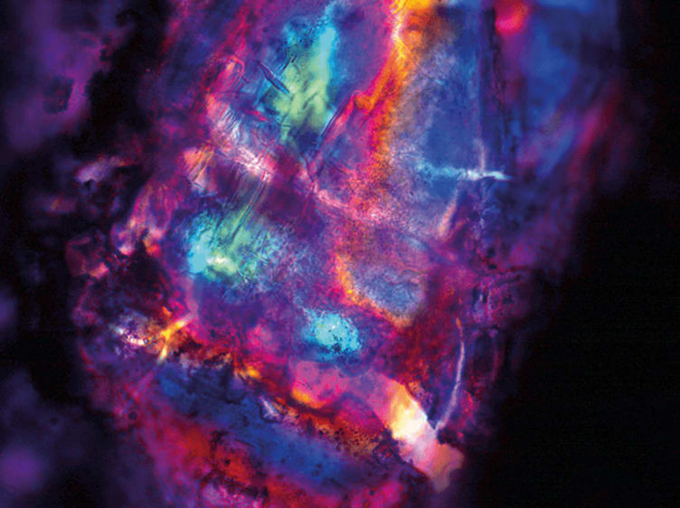
Microcosms Within a Crystalluria
This urinary cytology image was captured at 1000x by polarized light microscopy using an immersion objective and Papanicolaou stain.
JD Prieto, UGC Provincial de Anatomía Patológica and LABCO Pathology, Málaga, Spain.
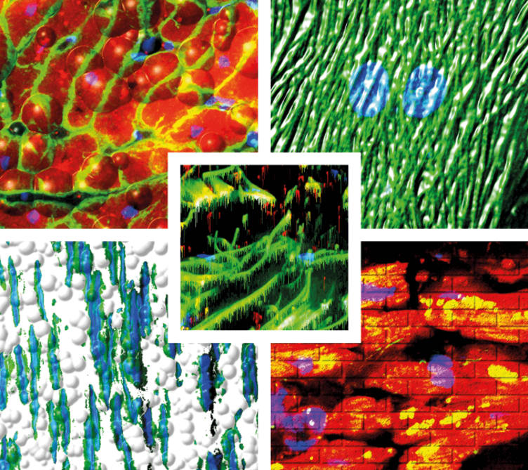
The Art of Fluorescence Deconvolution Imaging
A series of artistic images created using fluorescence deconvolution microscopy. Clockwise from top left: “Bubbling” dystrophin in heart; “plastic” cardiomyocytes; a “wall” of myocardium; smooth muscle “bubbles”; and “dripping” connective tissue in skin.
Brian J. Poindexter and Roger J. Bick, Multi-User Fluorescence Imaging and Microscopy Core Lab, UT McGovern Medical School, USA.
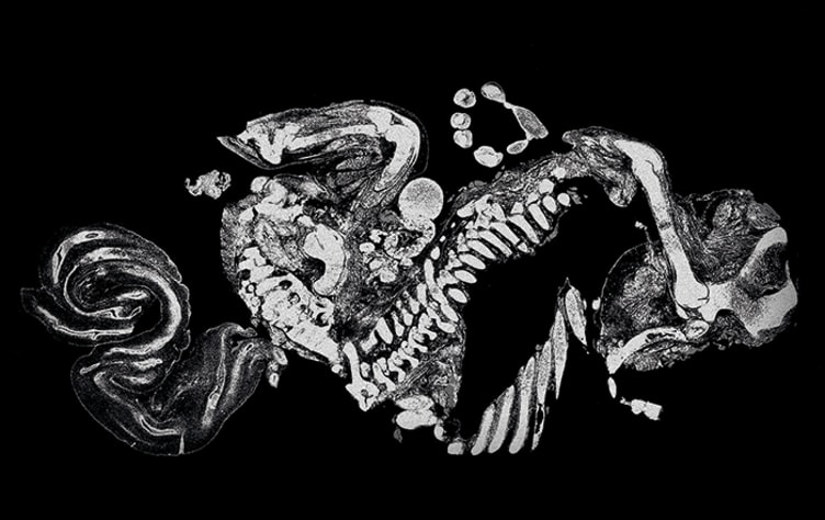
Parasagittal Section of Fetus
A whole slide image of a parasagittal section of the human fetus. Original magnification 0.45x.
Vamsi Parini, Loyola University Medical Center, USA.
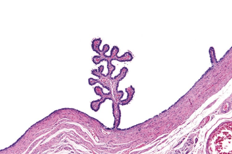
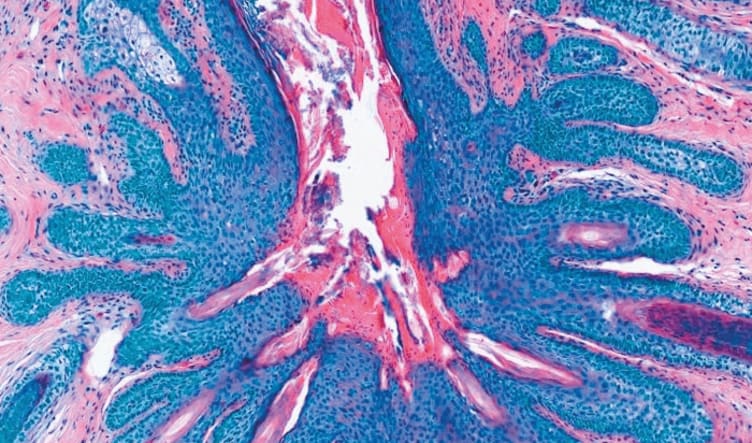
In the Desert of Paraffin Island
A lonely papillary projection found amidst tubal epithelia in a dilated hydrosalpinx specimen.
Marcelo Balancin, São Paulo, Brazil.
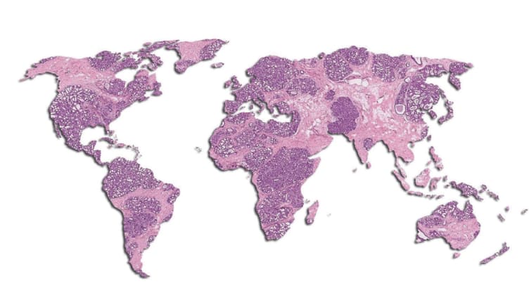
The Wide World of Histopathology
This world map offers a creative take on the vast scope of tissue pathology.
Nathan Swailes, @ihearthisto
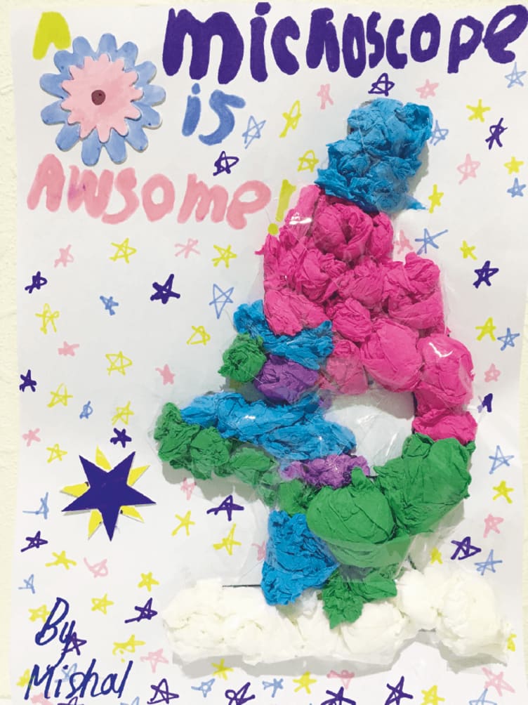
A Microscope is Awesome
This mixed-media piece was created by the nine-year-old daughter of a pathologist – perhaps a future pathologist herself!
Mishal Azam, Edgbaston High School for Girls, Birmingham, UK.
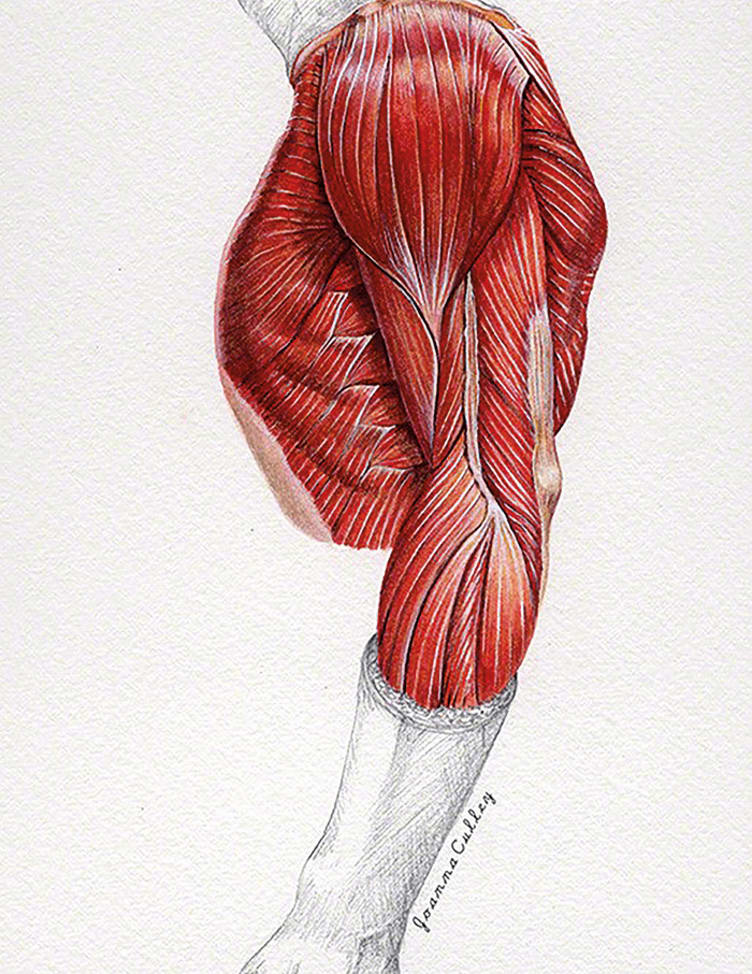
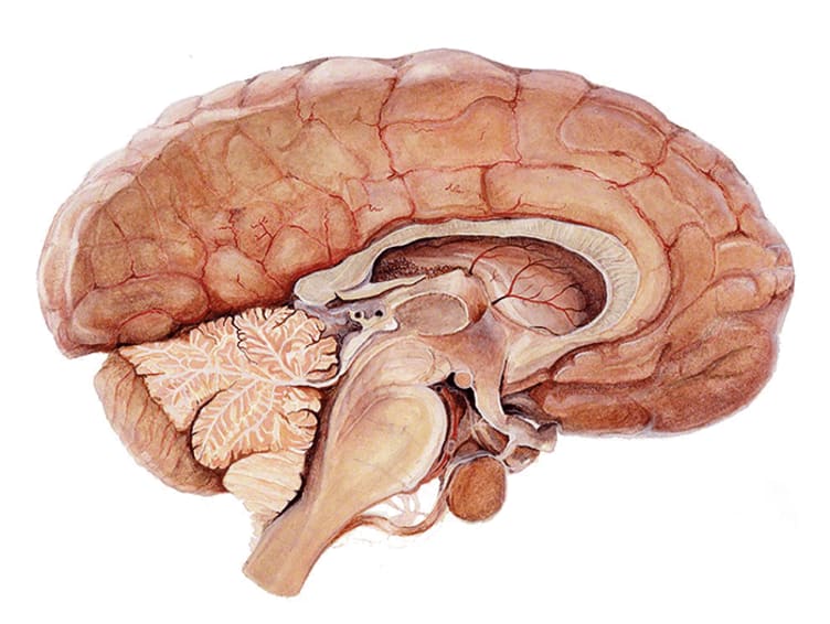
Works in Watercolor
Musculature of the arm and chest (top) and brain showing an enlarged pituitary gland (bottom).
Joanna Culley, Medical Artist, medical-artist.com

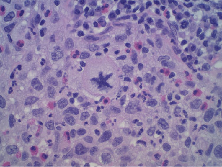
Starfish
An atypical mitotic figure identified in high-grade invasive urothelial carcinoma.
Craig Hart, York Pathology Associates LLC, USA.
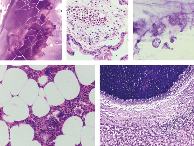
The Beauty of Pathology
Clockwise from top left: The cracked-glass appearance of a colloid; nucleated red blood cells in a placental villus; a hydatid daughter cyst; a calcified liver cyst; and a myelolipoma.
Sunil Swami, India.

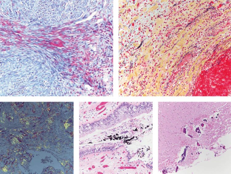
Death Under Glass
Clockwise from top left: Trichrome stain of muscle (blue: connective tissue, red: muscle fibers); pentachrome stain (black: nuclei and elastic fibers, yellow: collagen and reticular fibers, red: muscle, blue: mucin, bright red: fibrin); a shotgun blast to the head (pink: brain tissue, purple: bone); respiratory tract with soot; and amyloidosis, recognizable by its unique apple-green color.
Marianne Hamel and Nikki Johnson, The Death Under Glass Project, USA.

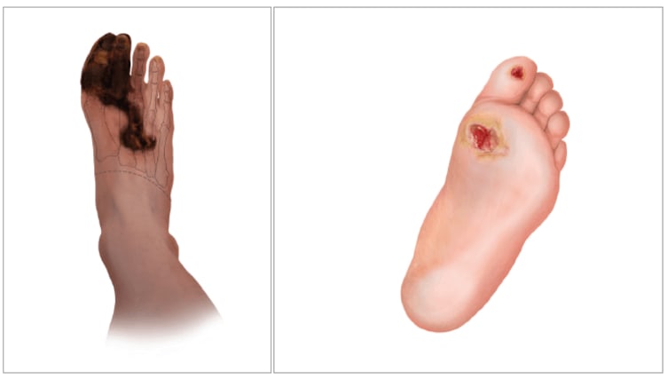
Podiatric Pathology
Wet and dry gangrene of the foot (left) and diabetic foot ulcers (right).
Merlin Strangeway, Medical Artists’ Association of Great Britain, merlinevans.com

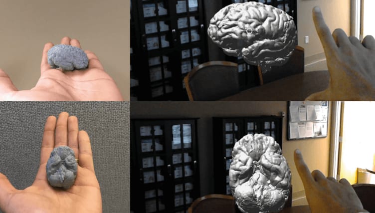
How Images Are Made
A composite of a human brain, both as a handheld 3D printout and displayed as a 3D hologram viewed in the Microsoft HoloLens.
Matthew Hanna and Liron Pantanowitz, University of Pittsburgh Medical Center, USA.

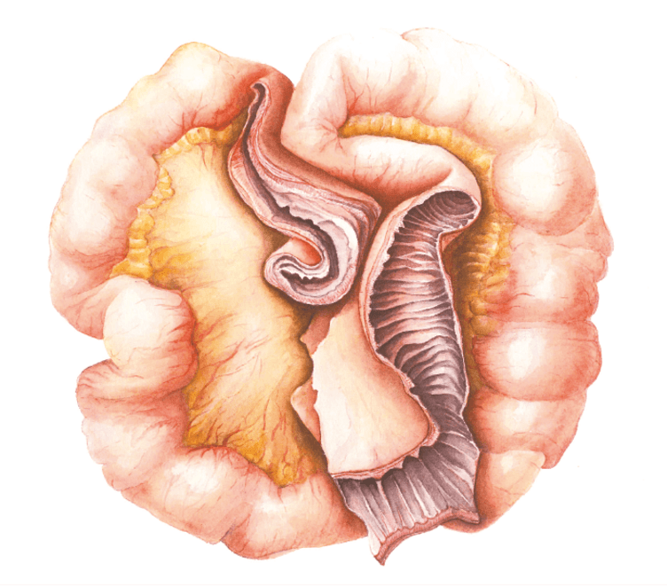
Radiation Structure of the Terminal Ileum
The wall of the ileum in this traditional watercolor illustration is grossly thickened and the lumen narrowed, with a healthy section also displayed to show the difference.
Louise Hinman, artistamedico.com
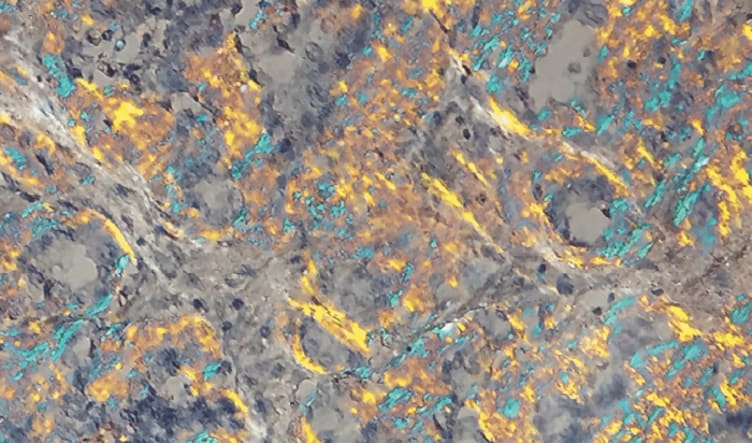
In a SFOG
Renal tubules in an acid-fuchsin orange G (SFOG) stain (left) and thyroid colloid (right).
Simone Münst, University Hospital Basel, Switzerland.

Cellularity Study #3
Acrylic on canvas.
Xiaoyin “Sara” Jiang, Duke University School of Medicine, USA.

Inside the Kidney
Membranous nephropathy.
Beth Braunhut, The University of Chicago, USA.

Trichofolliculoma
A trichofolliculoma (benign skin tumor) is composed of primitive hair follicles radiating from a central pore. In this image, the colors of the original H&E-stained tissue have been digitally altered for artistic effect.

Iron Marrow
In this image of bone marrow, the Prussian blue stain reveals the presence of iron.
Ziad El-Zaatari, Houston Methodist Hospital, USA

Abstract Pathology
Cytology preparation of a nipple discharge.
Darin L. Wolfe, Indiana Forensic & Surgical Pathology LLC, USA.

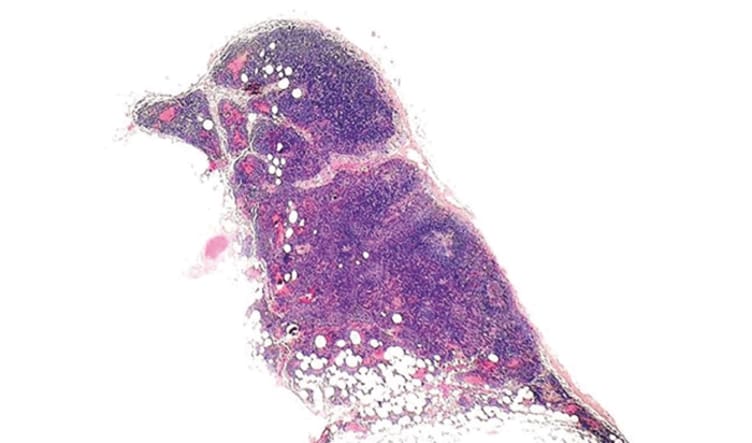
Sentinel Sparrow
“Aviary” nice example of a benign lymph node!
Katrina Collins, Hartford Hospital, USA.

Keep Rotating!
This is a high-power view of seminal vesicles showing amyloid depositions stained by Congo Red. Continue to play with your polarizer. You may encounter more beautiful colors than conventional apple-green birefringence!
Woo Cheal Cho, @DrAldehyde

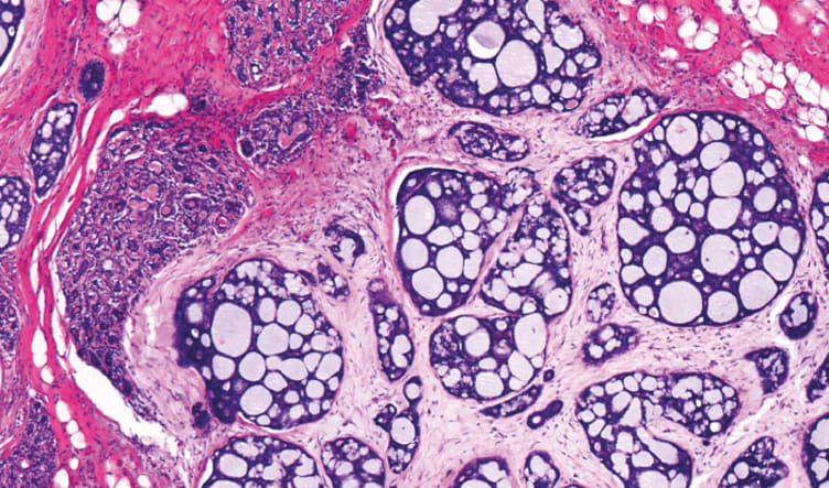
An ACCurate Diagnosis
Adenoid cystic carcinoma of the breast.
Dharam M. Ramnani, webpathology.com

Newsletters
Receive the latest pathologist news, personalities, education, and career development – weekly to your inbox.