The Pathologist
  • Explore Pathology

    Explore

    • Latest
    • Insights
    • Case Studies
    • Opinion & Personal Narratives
    • Research & Innovations
    • Product Profiles

    Featured Topics

    • Molecular Pathology
    • Infectious Disease
    • Digital Pathology

    Issues

    • Latest Issue
    • Archive
  • Subspecialties
    • Oncology
    • Histology
    • Cytology
    • Hematology
    • Endocrinology
    • Neurology
    • Microbiology & Immunology
    • Forensics
    • Pathologists' Assistants
  • Training & Education

    Career Development

    • Professional Development
    • Career Pathways
    • Workforce Trends

    Educational Resources

    • Guidelines & Recommendations
    • App Notes
    • eBooks

    Events

    • Webinars
    • Live Events
  • Events
    • Live Events
    • Webinars
  • Profiles & Community

    People & Profiles

    • Power List
    • Voices in the Community
    • Authors & Contributors
  • Multimedia
    • Video
    • Pathology Captures
Subscribe
Subscribe

False

The Pathologist / Issues / 2022 / Apr / Observing Infection
Microbiology & Immunology Microscopy and imaging Microbiology and Immunology

Observing Infection

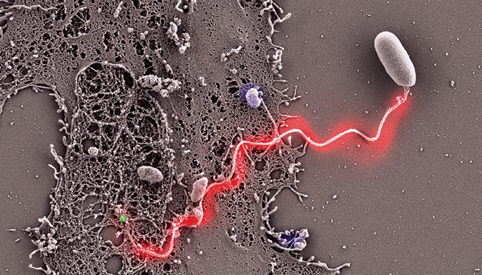
Correlative light and electron microscopy analysis of Encephalitozoon hellem infection of a host cell

04/12/2022 Gallery 1 min read

Share

Credit: Louis M. Weiss and Bing Han, Albert Einstein College of Medicine, Bronx, New York, USA. Image reprinted with permission from (1).

An Encephalitozoon hellem-infected host culture was incubated with rab-PcAb-EhPTP1 (rabbit polyclonal antibody to polar tube protein 1; red) and mAb-EhPTP4 (mouse monoclonal antibody to polar tube protein 4; green). The fluorescence and SEM images of the same site were taken sequentially, and the fluorescence images showing the labeling of EhPTP4 and the polar tube were correlated to the SEM images demonstrating germination of this E. hellem spore at high resolution. This image shows a germinated polar tube with EhPTP4 staining at the end of the tube and EhPTP1 staining along the length of the polar tube. Bar size: 2 μm.

Newsletters

Receive the latest pathologist news, personalities, education, and career development – weekly to your inbox.
Newsletter Signup Image

References

  1.  B Han et al., PLoS Pathog, 13, e1006341 (2017). PMID: 28426751.

Explore More in Pathology

Dive deeper into the world of pathology. Explore the latest articles, case studies, expert insights, and groundbreaking research.

False

Advertisement

Recommended

False

Affiliations:

Specialties:

Areas of Expertise:

Contributions:

False

The Pathologist
Subscribe

About

  • About Us
  • Work at Conexiant Europe
  • Terms and Conditions
  • Privacy Policy
  • Advertise With Us
  • Contact Us

Copyright © 2026 Texere Publishing Limited (trading as Conexiant), with registered number 08113419 whose registered office is at Booths No. 1, Booths Park, Chelford Road, Knutsford, England, WA16 8GS.