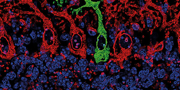
Most of us are familiar with liquid crystals as a technology involved in creating television and computer screens, but now, researchers at the University of Chicago’s Institute for Molecular Engineering have found a very different use for them – detecting amyloid fibrils, which are widely implicated in neurodegenerative diseases such as Alzheimer’s, Huntington’s and Parkinson’s (1), and are also suspected of playing a role in type 2 diabetes (2). Current methods for detecting amyloid fibrils are less than ideal – the fibers are thought to be at their most toxic early on in their formation, when they are still too small to study using optical microscopy. This means costly and complex techniques using fluorescence or neutron scattering are required.
Liquid crystals could provide a simple and relatively inexpensive way to study the tiny fibers, according to the University researchers. The method exploits the way the crystals respond to surface disturbances – a light-blocking layer is added to a film of liquid crystals, and on top of this a membrane, covered in water, is added. The molecules that form the toxic amyloid aggregates are injected into the water layer, and as the aggregates form and grow on the membrane, they imprint their shape into the liquid crystal underneath. This distorts the molecules present in the light-blocking layer, allowing light to shine through, explains co-author of the associated paper (3), Juan de Pablo. Though the fibers themselves are still too small to view, their effect is magnified by the imprint they leave, and is large enough to view using a simple optical microscope. “The liquid crystal is actually reporting what’s happening to the aggregates at the interface, and these bright spots become bigger and adopt the shape of the actual fibers that the protein is forming. Except you’re not seeing the fibers, you’re seeing the liquid crystal’s response to the fibers,” says de Pablo.
The team is now working on a way to use their method in emulsion as opposed to a flat surface, and they hope that eventually the technology could be used to create a method for testing small samples of blood or other body fluids, or to study the effects of different drugs on aggregate growth. “For research in type 2 diabetes, or Alzheimer’s or Parkinson’s, having this simple platform to perform these tests at a fraction of the cost of what’s required for fluorescence or neutron scattering would be very useful,” says de Pablo.
References
- M Stefani, “Structural features and cytotoxicity of amyloid oligomers: implications in Alzheimer’s disease and other diseases with amyloid deposits”, Prog Neurobiol, 99, 225–245 (2012). PMID: 22450705. A Mukherjee, et al., “Type 2 diabetes as a protein misfolding disease”, Trends Mol Med, 21, 439–449 (2015). PMID: 25998900. M Sadati, et al., “Liquid crystal enabled early stage detection of beta amyloid formation on lipid monolayers”, Adv Funct Mater, 25, 6050–6060 (2015).


