Techniques in Transition
Flexibility and computer-aided diagnosis can help digital pathology gain a foothold… but is the technology ready yet?
When it comes to “going digital,” pathologists fall into one of three groups: early adopters who are very enthusiastic, people in the middle who are unsure of its benefits, and those who are adamantly opposed to it (whose opinions range from skepticism to outright hostility). At the moment, I think it’s the early adopters who are driving the digital movement. That’s not to say that the skeptics are wrong – but pathologists are scientifically trained; they want to see evidence, and although there’s plenty of evidence to say that digital pathology is no worse than a light microscope, there’s no real proof that it’s better. It’s not an unreasonable view. What is needed is more evidence of a return on the investment required to go digital.
Return on investment
The first of these and most significant returns comes from the flexibility of workload distribution. That means convincing pathologists that they can report cases to hospitals other than their own. Once they buy into that, the traditional model for cellular pathology (where hospitals maintain their own laboratories with their own pathologists) breaks down completely. Instead, each hospital makes its own provision for pathology – and that might involve the use of remote reporting of sections via digital pathology. Why? Because some tasks are specialized, and it makes sense to have them performed by experts. Renal biopsies are a good example; it’s a complicated, small-volume area, requires some out of hours reporting and is very difficult for those who lack expertise – but if you are a renal pathologist it is feasible, and probably desirable to grow this workload by reporting renal biopsies for hospitals other than your own. By outsourcing this work, a hospital pathology department can concentrate on the volume specialties that it routinely deals with. Hospitals dealing with heavy workloads and often shorthanded through temporary or long term vacancies can use digital pathology to allow off-site reporting, either by locum pathologists or established consultants keen to work additional sessions. This avoids the need for travel, increases the pool of available expertise and can avoid expensive agency rates.
The other major promise of a return on investment is computer-aided diagnosis. There is very little of this actually in practice today but the potential is enormous. The spiraling demand for histopathology with our current aging population means histopathology needs increased automation and tools that improve pathologist’s decision-making ability. Automated slide review, mitotic counting, or tools that analyze biomarkers and grade tumors. These are all things that pathologists do every day, often not as well as we would like to think. We’re quite good at working out what’s abnormal – but our human eyes and brains struggle with objectively quantifying exactly how abnormal it is. So this is the next big thing in digital pathology… but it’s not ready yet. We don’t have the mitotic counting and tumor-grading algorithms we really need.
Algorithm development
There is some research around algorithm writing and development. There is not enough and it is limited in its extent. However, although showing some promise, these studies have not not been translated into practice for full-scale trials. There’s a much bigger step than most people realize in translating a written algorithm into a piece of software for diagnostic workstations, and most research teams don’t have the resource to do this, and many institutions active in this arena lack partnership with pathology labs with the IT infrastructure to support this sort of translation.
Our team at the newly established UHCW Centre of excellence for digital pathology are working on tumor grading. We’ve broken it down into the same parameters that are used for breast cancer grading – mitotic rate, nuclear pleomorphism, and degree of tubular differentiation in adenocarcinoma – and modeled each element with an algorithm. We’ve had some success, but the difficulty lies in getting good enough results from each parameter. The results thus far are definitely showing efficacy, but at some point we need to draw line, stop improving the algorithm, translate it, and evaluate how it actually works in a full-scale clinical trial. That will be an expensive step, but a valuable one – and one we hope to take in the next year and a half.
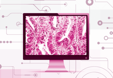
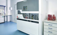
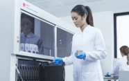
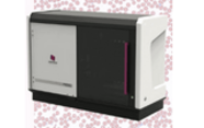
One thing we’ve learned is that scanning still has a long way to go. Modern-day scanners work quite well, but they have quite variable focus. They can focus most of a slide well enough for a pathologist to make sense of it, but that isn’t good enough for algorithms. Algorithms will not work on areas that aren’t perfectly in focus. So part of our current work is on a program designed to measure the slides’ degree of focus. That way, we can feed in only the appropriate cases and increase our success rate dramatically. Improving scan focus quality control is a key element to getting algorithms to work successfully in real-time.
You’ve also got to take into account where the algorithm is designed to act. Some will operate upstream of the pathologist, like a rare event detection tool we’re developing to find melanoma in sentinel node biopsies. The slides will be exported from the scanner to a server, where the algorithm runs, finds the area of interest, and the annotated slides are then returned to the pathologist reporting pool for review. That algorithm, which is designed to work on a server in the cloud and process massive numbers of slides from multiple centers, places special demands on the technology, which requires solving prior to roll out. You need software to sort slides at the source, send them to the cloud, bring them back with annotations visible on multiple viewing platforms with complete resilience and accuracy, and it has to be secure. None of this is new to the industry but much of it is to histopathology so we need to learn and catch up.
Other algorithms work downstream of the pathologist. That is, the pathologist identifies the slide’s area of interest and then passes it on to the algorithm – like in ER staining in breast cancer, where the tumor is manually identified at diagnosis and where estrogen receptor, progesterone receptor and HER2 are stained on sequential sections but scored according to the originally identified region of interest. These algorithms combine overlay steps identifying the region to be scored and then the scoring itself. Still others will work under the supervision of the pathologist, such as tumor grading tools performed at the point of the diagnosis. These will need to run on the workstation in real-time to allow the minimum of disruption to workflow.
Therefore, what the algorithm is doing places some demands on the way it needs to be designed from the ground up. You can’t do this later in the process; it has to be envisioned right at the start so that you can predict your software requirements. It’s not something most people think about, though. It’s only when you start to work with algorithms that you realize how much these things matter. And only when you find ways of addressing those issues can digital technologies truly support the work of pathologists.
The big picture
The only sensible reason to change to digital at the moment is to provide flexibility for slide reporting. At least in the United Kingdom, we just don’t have enough pathologists right now. Digital pathology offers a theoretical solution to that problem – we can reorganize ourselves as digital hubs and redistribute our slides so that we get better efficiency from the reporting manpower that we already have.
To administrators, I would say, “You’ve got to think big.” think about creating hub-and-spoke laboratory models and setting up networks to address the deficits caused by a lack of personnel. Digital pathology can solve many of the problems with those kinds of transitions – and that’s a major driver, because at least half of pathology labs in the UK right now are severely understaffed. If you give existing pathologists the work they do best, allowing them to properly subspecialize, rather than work on what has got to be done because there is no one else to do it, they work faster and more efficiently. I also suspect that it would reduce error. Error is very expensive in pathology, so the better you are at getting the answer right – which means giving pathologists the work at which they excel – the more costs, and lives, you can save.
In a nutshell...
- The greatest benefit of digital pathology lies in increased flexibility, which can help balance out the widespread shortage of pathologists
- Computer-aided diagnosis may also provide benefits one day – but the algorithms aren’t quite ready yet
Professor David Snead is Clinical Lead for cellular pathology at the University Hospitals Coventry and Warwickshire NHS Trust and Coventry and Warwickshire Pathology Services. He is the Director of the UHCW Centre of Excellence for Digital Pathology and Professor of Practice at Warwick Medical School, UK.
Read more:
The Doubting Dollar
By Luke Perkocha
A Roadmap to the Future
By Alexi Baidoshvili
Part of a Larger Whole
By Marcial García Rojo
A Business Case for Common Sense
By Liron Pantanowitz

While obtaining degrees in biology from the University of Alberta and biochemistry from Penn State College of Medicine, I worked as a freelance science and medical writer. I was able to hone my skills in research, presentation and scientific writing by assembling grants and journal articles, speaking at international conferences, and consulting on topics ranging from medical education to comic book science. As much as I’ve enjoyed designing new bacteria and plausible superheroes, though, I’m more pleased than ever to be at Texere, using my writing and editing skills to create great content for a professional audience.


