The Core of the Problem
Next generation tissue microarrays overcome punching inaccuracies and bring the technique into the era of precision medicine
At a Glance
- Tissue microarrays (TMAs) are increasingly used for biomarker evaluation and validation
- Conventional TMAs lack accuracy in punching, meaning that some areas of interest are captured inadequately or not at all
- Next generation TMAs (ngTMAs) are digitally scanned, annotated and cored, resulting in more precise alignment and better coring to optimize research
- Now that we’ve achieved precision coring, the next step for ngTMAs is to multiplex antibodies and improve image analysis
As the era of precision medicine dawns, the role of the pathologist is extending beyond diagnosis to include the interpretation of molecular tissue biomarkers. The ability to detect protein, RNA and DNA alterations in cancer now helps us to tailor prognoses and predict therapy responses. As a consequence, biomarker research has exploded over recent years – and with it, the number of studies using tissue microarrays (TMAs) as a tool for biomarker evaluation and validation. Essentially, the TMA is a tissue archive constructed by transferring small tissue cores, typically 0.6 or 1.0 mm in diameter, from a “donor” tissue block into a “recipient” block. Repeated multiple times, a recipient TMA block can carry up to 500 different tissue cores from multiple tumors or patients (Figure 1).
In 1998, Kononen et al. (1) published the first report using TMAs for high-throughput molecular profiling of cancers; at the same time, the report highlighted some of the technique’s great advantages. First and foremost, the high-throughput nature of the approach means that studies on a large number of patient tissues can be assembled on just a handful of TMA blocks. This can substantially reduce the cost of consumables, and can help prevent the depletion of tissue that should remain in the diagnostic archive for eventual re-evaluation. Staining procedures can be carried out simultaneously across different cores, eliminating experimental variability. Dozens of different biomarkers can be applied to serial sections of the TMA, ensuring its viability as a longstanding research tool for many study groups. Attributes like these mean that TMAs have become a mainstay in translational research.
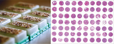
Figure 1. Left: a set of ngTMA blocks just after construction. Right: part of a large ngTMA with H&E staining.
What is the problem?
On the other hand, conventional tissue microarraying has always presented one major hurdle: the inaccuracy of punching. Typically, a region of interest is marked on the H&E slide, indicating that a core should be punched out from the encircled area. But when the tissues are cored out, there’s no physical alignment of the block and slide. This imposes a major limitation on biomarker research, because some specific targets exist only in certain histological areas and aren’t captured adequately – or at all. There have been no major developments in tissue microarraying since its inception, but now, with the substantial strides made in digital pathology, the technique is primed to evolve. At the Institute of Pathology of the University of Bern, TMAs are designed based on a next generation tissue microarray (ngTMA) approach that relies on a process we call “VDA2”: visualization, digitization, automation and analysis.
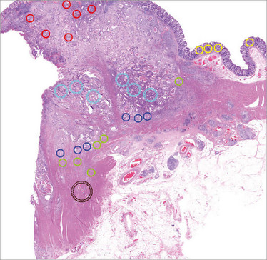
Figure 2. Scan of an H&E stain from a colorectal cancer. Digital annotations of different sizes and colors indicate the regions of interest. The next step in the ngTMA process is alignment of the annotated scan with a donor block image. Annotated regions are then cored and an ngTMA can be constructed.
Visualization
With ngTMA, our vision is to answer targeted research questions by optimally visualizing and evaluating biomarkers in tissue. Each ngTMA is designed with a specific purpose in mind – so each one is unique and allows planning for special considerations like tumor heterogeneity. Determining which specific histological interfaces are relevant for the study will help to determine the core size that should be used for punching the regions selected, as well as the number of cores per tissue sample. The number of biomarkers to be investigated will determine how many sections need to be cut and provide information on how many ngTMAs should be constructed. Consideration is also given to power and downstream statistical analysis – meaning that, no matter the experiment, we can design ngTMAs that efficiently deliver the necessary results.
Digitization
The integration of digital pathology into the TMA workflow opens new dimensions for biomarker research. Conventional (H&E or immunostained) histological slides can now be scanned and then annotated using a digital slide viewer. We can paste any number of TMA annotations, in various colors and sizes (0.6, 1.0, 1.5 or 2.0 mm in diameter), onto the scanned slide and move them to our histological region of interest (Figure 2). We can then align an image of each donor block to the digital scan containing these annotations and use their coordinates for precise coring and ngTMA construction. Annotations can be placed in any area of the digital slide, and the precision of the alignment between block and slide guarantees that the marked spot is cored out correctly.
Automation
We can core blocks using an automated tissue microarrayer. As such, the speed and convenience with which we can generate TMAs is far beyond that of conventional techniques. For example, three TMA blocks containing 475 spots each would take approximately 84 hours to construct using a homemade or semi-automated device, but under three hours with an automated arrayer. The automated approach also minimizes loss of tissue cores and maps each core to a particular position within the TMA grid, so the risk of error or spot confusion is negligible. Actual images of the cored donor blocks and their corresponding annotations can also be taken, ensuring traceability of each TMA spot back to the original block for quality assurance.
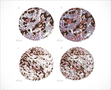
Figure 3. Digital image analysis of an ngTMA that has undergone double immunohistochemistry for pan-cytokeratin (brown) and CD68 (red). This image shows CD68+ macrophages at the invasion front of a colorectal cancer.
Analysis
Combined with digital image analysis, ngTMA is a powerful tool. It allows researchers to generate objective and reproducible data to help fast-track biomarker validation, and it can be used as an investigative tool to further unmask the biology of cancers.
An excellent example to showcase the power of the combined ngTMA/digital image analysis approach is the tumor microenvironment of colorectal cancers. The tumor microenvironment is emerging as a highly relevant histological area for the identification of novel prognostic and predictive biomarkers. Not only T cells, but also macrophages, fibroblasts, and other stromal cells are known to play pro- or anti-tumor roles associated with better or worse patient outcomes (2,3) (Figure 3). In fact, depending on the context, some cell types may have both pro- and anti-tumor functions – meaning that the number of biomarkers required to help tweeze out these contributions and subclassify cell populations is growing. The region at the tumor front where the most invasive cells meet the tumor stroma holds a wealth of potential information we still need to measure, quantify and ultimately use. Recently, the presence of tumor budding was shown to be an adverse prognostic factor, but also that the balance between attacker (tumor buds) and defender (CD8+ T cells) may have a more relevant clinical impact than either feature alone (4) (Figure 4).
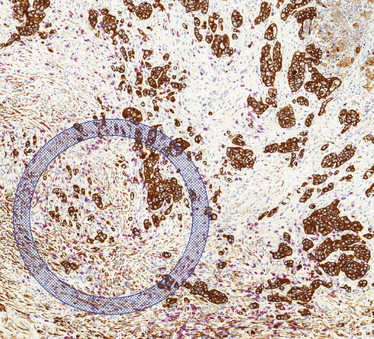
Figure 4. Scan with ngTMA annotation of a colorectal cancer, highlighting a dense area of tumor budding (pan-cytokeratin; brown) and surrounding CD8+ lymphocytes (red). The annotated region can be cored out and included into an ngTMA.
How can we get the most information out of these clearly relevant findings? ngTMAs of the tumor microenvironment, combined with digital analysis, will help unravel the contributions of different cell types to tumor progression, metastasis and ultimately clinical outcome. Pattern recognition can be used to identify different structures at the invasion front. We can even measure staining intensities and set thresholds for positive staining. Three-dimensional reconstruction of the invasion front will help determine the spatial distribution between different cells and structures and even identify apparent contact between cell types. These features supplement “standard” image analysis like quantifying the number of positive cells within a region of interest or separating by color, size, shape or other morphological or cytological features.
What does the future of ngTMA hold?
Areas like the tumor microenvironment highlight our need for multiplexing. Although we can now capture precise histological areas using ngTMA, we are still limited by our ability to multiplex various antibodies or probes, especially using chromogenic methods. Not only should we be able to do this in the lab, but image analysis should ideally allow us to separate out these components from actual immunostained slides. It’s true that this kind of in-depth work will send the amount of data we produce skyrocketing – so it will become increasingly important to work out the best ways of streamlining our analyses and sharing them with other researchers.
ngTMA is an opportunity to take biomarker research to a new level in clinical and translational settings. Because it’s a method that combines histopathological expertise with targeted research questions, the latest digital pathology technology, and automated arraying, ngTMA is a way of achieving better characterization and validation of biomarkers – and one I expect will continue to grow in popularity.
- J Kononen et al., “Tissue microarrays for highthroughput molecular profiling of tumor pecimens”, Nat Med, 4, 844–847 (1998). PMID: 9662379.
- C Isella et al., “Stromal contribution to the colorectal cancer transcriptome”, Nat Genet, 47, 312–319 (2015). PMID: 25706627.
- J Galon et al., “Type, density, and location of immune cells within human colorectal tumors predict clinical outcome”, Science, 313, 1960–1964 (2006). PMID: 17008531.
- A Lugli et al., “CD8+ lymphocytes/tumour-budding index: an independent prognostic factor representing a ‘pro-/anti-tumour’ approach to tumour host interaction in colorectal cancer”, Br J Cancer, 101, 1382–1392 (2009). PMID: 19755986.
Inti Zlobec is Head of the Translational Research Unit (TRU) at the Institute of Pathology, University of Bern, Switzerland.

