Taking Aim at a Moving Target
A new imaging technique allows us to see the evolution of cancerous cells
How do cancerous tumors form? It’s a question to which we don’t yet have a detailed answer – but one that, if fully explored, could lead to better prevention, diagnosis, and treatment. Using a laboratory model of breast cancer, a team of researchers at the Walter and Eliza Hall Institute of Medical Research in Parkville, Australia, has developed a new imaging technique to observe individual cells within a tumor at high resolution and in three dimensions (1). The approach allows them to observe individual clones descended from a single precancerous cell to see how they change, which become cancerous, and which ones are most changeable or have potential growth or treatment resistance advantages. We spoke to study author Jane Visvader to learn more…
What enabled you to view breast cancer tumors in 3D at such a resolution?
The clearing agent was instrumental in enabling us to simultaneously perform immunolabeling and detect native fluorescence in large portions of tissue or tumors, and in allowing us to gain insight into tissue architecture at single cell resolution.
What's the most interesting discovery revealed by your new imaging technique?
The pipeline involving 3D imaging at high resolution, together with the sorting and sequencing of individual clones, led to the unexpected finding that almost every clone had undergone an epithelial-to-mesenchymal transition (EMT). Although this process has been well-documented in cancers, it was presumed to occur in relatively rare cells, such as those along the leading edge of the tumor, before their extravasation into the bloodstream. In the p53/Pten deletion models we examined, the EMT was widespread and occurred at a clonal level. Of note, the molecular signature changed drastically from an epithelial to mesenchymal state, based not just on a small number of genes, but on hundreds to thousands.
These findings highlight the inherent plasticity of tumor cells and indicate that both epithelial and mesenchymal cells need to be targeted early in tumor progression. It is somewhat alarming that each clone had a propensity to shift towards a different (mesenchymal) state, indicating that cancer cells behave like moving targets.
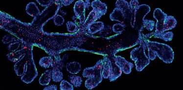
Credit: Rios, Visvader et al., published in Cancer Cell
What’s next for this research?
Robust markers of the epithelial, mesenchymal, and hybrid cancerous states must be identified to allow more effective targeting – but this is a tough proposition and will require further research.
As it stands, our imaging protocol is simple to use, relies on a non-toxic clearing agent, and can be completed within three days. My colleagues and I hope that it will one day be adapted for use in pathology labs to diagnose and select treatment for specific diseases.
- AC Rios et al., “Intraclonal plasticity in mammary tumors revealed through large-scale single-cell resolution 3D imaging”, Cancer Cell, 35, 953 (2019). PMID: 31185217.

While obtaining degrees in biology from the University of Alberta and biochemistry from Penn State College of Medicine, I worked as a freelance science and medical writer. I was able to hone my skills in research, presentation and scientific writing by assembling grants and journal articles, speaking at international conferences, and consulting on topics ranging from medical education to comic book science. As much as I’ve enjoyed designing new bacteria and plausible superheroes, though, I’m more pleased than ever to be at Texere, using my writing and editing skills to create great content for a professional audience.


